 Movie
Movie Controller

[English] 日本語
 Yorodumi
- PDB-8trc: mGluR3 in the presence of the antagonist LY 341495 and positive a... -
+ Open data

- Basic information
| Entry | Database: PDB / ID: 8trc | ||||||||||||
|---|---|---|---|---|---|---|---|---|---|---|---|---|---|
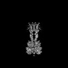
| Title | mGluR3 in the presence of the antagonist LY 341495 and positive allosteric modulator VU6023326 | ||||||||||||
 Components Components | Metabotropic glutamate receptor 3 | ||||||||||||
 Keywords Keywords | MEMBRANE PROTEIN / GPCR / synaptic protein | ||||||||||||
| Function / homology |  Function and homology information Function and homology informationClass C/3 (Metabotropic glutamate/pheromone receptors) / group II metabotropic glutamate receptor activity / G protein-coupled glutamate receptor signaling pathway / G alpha (i) signalling events / astrocyte projection / cellular response to stress / postsynaptic modulation of chemical synaptic transmission / regulation of synaptic transmission, glutamatergic / sensory perception of pain / calcium channel regulator activity ...Class C/3 (Metabotropic glutamate/pheromone receptors) / group II metabotropic glutamate receptor activity / G protein-coupled glutamate receptor signaling pathway / G alpha (i) signalling events / astrocyte projection / cellular response to stress / postsynaptic modulation of chemical synaptic transmission / regulation of synaptic transmission, glutamatergic / sensory perception of pain / calcium channel regulator activity / modulation of chemical synaptic transmission / adenylate cyclase-inhibiting G protein-coupled receptor signaling pathway / gene expression / presynaptic membrane / scaffold protein binding / dendritic spine / postsynaptic membrane / neuron projection / postsynaptic density / axon / glutamatergic synapse / plasma membrane Similarity search - Function | ||||||||||||
| Biological species |  | ||||||||||||
| Method | ELECTRON MICROSCOPY / single particle reconstruction / cryo EM / Resolution: 3.3 Å | ||||||||||||
 Authors Authors | Strauss, A. / Levitz, J. | ||||||||||||
| Funding support |  United States, 3items United States, 3items
| ||||||||||||
 Citation Citation | Journal: bioRxiv / Year: 2023 Title: Structural basis of allosteric modulation of metabotropic glutamate receptor activation and desensitization. Abstract: The metabotropic glutamate receptors (mGluRs) are neuromodulatory family C G protein coupled receptors which assemble as dimers and allosterically couple extracellular ligand binding domains (LBDs) ...The metabotropic glutamate receptors (mGluRs) are neuromodulatory family C G protein coupled receptors which assemble as dimers and allosterically couple extracellular ligand binding domains (LBDs) to transmembrane domains (TMDs) to drive intracellular signaling. Pharmacologically, mGluRs can be targeted either at the LBDs by glutamate and synthetic orthosteric compounds or at the TMDs by allosteric modulators. Despite the potential of allosteric TMD-targeting compounds as therapeutics, an understanding of the functional and structural basis of their effects on mGluRs is limited. Here we use a battery of approaches to dissect the distinct functional and structural effects of orthosteric versus allosteric ligands. We find using electrophysiological and live cell imaging assays that both agonists and positive allosteric modulators (PAMs) can drive activation and desensitization of mGluRs. The effects of PAMs are pleiotropic, including both the ability to boost the maximal response to orthosteric agonists and to serve independently as desensitization-biased agonists across mGluR subtypes. Conformational sensors reveal PAM-driven inter-subunit re-arrangements at both the LBD and TMD. Motivated by this, we determine cryo-electron microscopy structures of mGluR3 in the presence of either an agonist or antagonist alone or in combination with a PAM. These structures reveal PAM-driven re-shaping of intra- and inter-subunit conformations and provide evidence for a rolling TMD dimer interface activation pathway that controls G protein and beta-arrestin coupling. HIGHLIGHTS: -Agonists and PAMs drive mGluR activation, desensitization, and endocytosis-PAMs are desensitization-biased and synergistic with agonists-Four combinatorial ligand conditions reveal an ...HIGHLIGHTS: -Agonists and PAMs drive mGluR activation, desensitization, and endocytosis-PAMs are desensitization-biased and synergistic with agonists-Four combinatorial ligand conditions reveal an ensemble of full-length mGluR structures with novel interfaces-Activation and desensitization involve rolling TMD interfaces which are re-shaped by PAM. | ||||||||||||
| History |
|
- Structure visualization
| Structure viewer | Molecule: MolmilJmol/JSmol |
|---|
- Downloads & links
-Download
| PDBx/mmCIF format | 8trc.cif.gz | 292.2 KB | Display | PDBx/mmCIF format |
|---|---|---|---|---|
| PDB format | pdb8trc.ent.gz | 187.8 KB | Display | PDB format |
| PDBx/mmJSON format | 8trc.json.gz | Tree view | PDBx/mmJSON format | |
| Others |  Other downloads Other downloads |
-Validation report
| Arichive directory | https://data.pdbj.org/pub/pdb/validation_reports/tr/8trcftp://data.pdbj.org/pub/pdb/validation_reports/tr/8trc | HTTPS FTP |
|---|
-Related structure data
| Related structure data |  41577MC 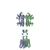 8tqbC 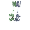 8tr0C 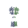 8tr2C M: map data used to model this data C: citing same article ( |
|---|---|
| Similar structure data |


-Links
 PDBj
PDBj


- Assembly
| Deposited unit | 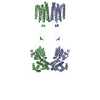
|
|---|---|
| 1 |
|
-Components
| #1: Protein | Mass: 103161.555 Da / Num. of mol.: 2 Source method: isolated from a genetically manipulated source Source: (gene. exp.)  Homo sapiens (human) / References: UniProt: P31422 Homo sapiens (human) / References: UniProt: P31422#2: Chemical |   Mass: 353.369 Da / Num. of mol.: 2 / Source method: obtained synthetically / Formula: C20H19NO5 / Feature type: SUBJECT OF INVESTIGATION Mass: 353.369 Da / Num. of mol.: 2 / Source method: obtained synthetically / Formula: C20H19NO5 / Feature type: SUBJECT OF INVESTIGATIONHas ligand of interest | Y | Has protein modification | Y | |
|---|
-Experimental details
-Experiment
| Experiment | Method: ELECTRON MICROSCOPY |
|---|---|
| EM experiment | Aggregation state: PARTICLE / 3D reconstruction method: single particle reconstruction |
- Sample preparation
| Component | Name: Metabotropic Glutamate Receptor 3 dimer / Type: COMPLEX / Entity ID: #1 / Source: RECOMBINANT | |||||||||||||||||||||
|---|---|---|---|---|---|---|---|---|---|---|---|---|---|---|---|---|---|---|---|---|---|---|
| Molecular weight | Value: 0.26 MDa / Experimental value: NO | |||||||||||||||||||||
| Source (natural) | Organism: | |||||||||||||||||||||
| Source (recombinant) | Organism: Homo sapiens (human) | |||||||||||||||||||||
| Buffer solution |
| |||||||||||||||||||||
| Specimen |
| |||||||||||||||||||||
| Specimen support |
| |||||||||||||||||||||
| Vitrification |
|
- Electron microscopy imaging
| Experimental equipment |  Model: Titan Krios / Image courtesy: FEI Company |
|---|---|
| Microscopy | Model: FEI TITAN KRIOS |
| Electron gun | Electron source:  FIELD EMISSION GUN / Accelerating voltage: 300 kV / Illumination mode: FLOOD BEAM FIELD EMISSION GUN / Accelerating voltage: 300 kV / Illumination mode: FLOOD BEAM |
| Electron lens | Mode: BRIGHT FIELD / Nominal defocus max: 2000 nm / Nominal defocus min: 700 nm / Alignment procedure: BASIC |
| Specimen holder | Cryogen: NITROGEN / Specimen holder model: FEI TITAN KRIOS AUTOGRID HOLDER |
| Image recording | Electron dose: 58 e/Å2 / Detector mode: COUNTING / Film or detector model: GATAN K3 BIOQUANTUM (6k x 4k) |
- Processing
| CTF correction | Type: PHASE FLIPPING AND AMPLITUDE CORRECTION |
|---|---|
| 3D reconstruction | Resolution: 3.3 Å / Resolution method: FSC 0.143 CUT-OFF / Num. of particles: 116376 / Symmetry type: POINT |
| Atomic model building | Protocol: AB INITIO MODEL / Space: REAL |
