 Movie
Movie Controller


+ Open data

- Basic information
| Entry | Database: PDB / ID: 8tdi | ||||||
|---|---|---|---|---|---|---|---|
| Title | Structure of P2B11 Glucuronide-3-dehydrogenase | ||||||
 Components Components | P2B11 Glucuronide-3-dehydrogenase | ||||||
 Keywords Keywords | OXIDOREDUCTASE / Dehydrogenase | ||||||
| Function / homology | NICOTINAMIDE-ADENINE-DINUCLEOTIDE Function and homology information Function and homology information | ||||||
| Biological species |  Collinsella tanakaei (bacteria) Collinsella tanakaei (bacteria) | ||||||
| Method |  X-RAY DIFFRACTION / SYNCHROTRON / MOLECULAR REPLACEMENT / Resolution: 2.6 Å X-RAY DIFFRACTION / SYNCHROTRON / MOLECULAR REPLACEMENT / Resolution: 2.6 Å | ||||||
 Authors Authors | Lazarski, A.C. / Worrall, L.J. / Strynadka, N.C.J. | ||||||
| Funding support |  Canada, 1items Canada, 1items
| ||||||
 Citation Citation | Journal: Nature / Year: 2024 Title: An alternative broad-specificity pathway for glycan breakdown in bacteria. Authors: Nasseri, S.A. / Lazarski, A.C. / Lemmer, I.L. / Zhang, C.Y. / Brencher, E. / Chen, H.M. / Sim, L. / Panwar, D. / Betschart, L. / Worrall, L.J. / Brumer, H. / Strynadka, N.C.J. / Withers, S.G. | ||||||
| History |
|
- Structure visualization
| Structure viewer | Molecule: MolmilJmol/JSmol |
|---|
- Downloads & links
-Download
| PDBx/mmCIF format | 8tdi.cif.gz | 885.5 KB | Display | PDBx/mmCIF format |
|---|---|---|---|---|
| PDB format | pdb8tdi.ent.gz | 739.7 KB | Display | PDB format |
| PDBx/mmJSON format | 8tdi.json.gz | Tree view | PDBx/mmJSON format | |
| Others |  Other downloads Other downloads |
-Validation report
| Arichive directory | https://data.pdbj.org/pub/pdb/validation_reports/td/8tdiftp://data.pdbj.org/pub/pdb/validation_reports/td/8tdi | HTTPS FTP |
|---|
-Related structure data
| Related structure data |  8tcdC  8tcrC 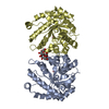 8tcsC  8tctC  8tdaC  8tdeC  8tdfC 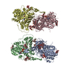 8tdhC  8v31C C: citing same article ( |
|---|---|
| Similar structure data |
-Links
 PDBj
PDBj- Assembly
| Deposited unit | 
| ||||||||
|---|---|---|---|---|---|---|---|---|---|
| 1 | 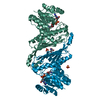
| ||||||||
| 2 | 
| ||||||||
| 3 | 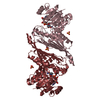
| ||||||||
| 4 | 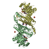
| ||||||||
| 5 | 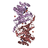
| ||||||||
| 6 | 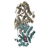
| ||||||||
| Unit cell |
|
-Components
| #1: Protein | Mass: 45297.680 Da / Num. of mol.: 12 Source method: isolated from a genetically manipulated source Source: (gene. exp.) Collinsella tanakaei (bacteria) / Production host: #2: Chemical | ChemComp-NAD /   Mass: 663.425 Da / Num. of mol.: 12 / Source method: obtained synthetically / Formula: C21H27N7O14P2 / Comment: NAD*YM Mass: 663.425 Da / Num. of mol.: 12 / Source method: obtained synthetically / Formula: C21H27N7O14P2 / Comment: NAD*YM#3: Chemical | ChemComp-SO4 /   Mass: 96.063 Da / Num. of mol.: 14 / Source method: obtained synthetically / Formula: SO4 Mass: 96.063 Da / Num. of mol.: 14 / Source method: obtained synthetically / Formula: SO4#4: Chemical | ChemComp-TRS /   Mass: 122.143 Da / Num. of mol.: 8 / Source method: obtained synthetically / Formula: C4H12NO3 / Comment: pH buffer*YM Mass: 122.143 Da / Num. of mol.: 8 / Source method: obtained synthetically / Formula: C4H12NO3 / Comment: pH buffer*YM#5: Water | ChemComp-HOH / |  Mass: 18.015 Da / Num. of mol.: 245 / Source method: isolated from a natural source / Formula: H2O Mass: 18.015 Da / Num. of mol.: 245 / Source method: isolated from a natural source / Formula: H2OHas ligand of interest | N | |
|---|
-Experimental details
-Experiment
| Experiment | Method: X-RAY DIFFRACTION / Number of used crystals: 1 |
|---|
- Sample preparation
| Crystal | Density Matthews: 2.46 Å3/Da / Density % sol: 50.04 % |
|---|---|
| Crystal grow | Temperature: 293 K / Method: vapor diffusion, sitting drop Details: 0.2 M LiSO4 19% PEG 3350 0.1 M Bis-Tris methane pH 5.5 |
-Data collection
| Diffraction | Mean temperature: 100 K / Serial crystal experiment: N |
|---|---|
| Diffraction source | Source: SYNCHROTRON / Site: CLSI / Beamline: 08B1-1 / Wavelength: 1.18 Å |
| Detector | Type: DECTRIS PILATUS3 S 6M / Detector: PIXEL / Date: Apr 14, 2023 |
| Radiation | Protocol: SINGLE WAVELENGTH / Monochromatic (M) / Laue (L): M / Scattering type: x-ray |
| Radiation wavelength | Wavelength: 1.18 Å / Relative weight: 1 |
| Reflection | Resolution: 2.6→48.37 Å / Num. obs: 161121 / % possible obs: 100 % / Redundancy: 6.9 % / CC1/2: 0.999 / Rmerge(I) obs: 0.121 / Rpim(I) all: 0.049 / Rrim(I) all: 0.13 / Χ2: 1.02 / Net I/σ(I): 13.7 / Num. measured all: 1108051 |
| Reflection shell | Resolution: 2.6→2.64 Å / % possible obs: 99.9 % / Redundancy: 7 % / Rmerge(I) obs: 1.919 / Num. measured all: 55713 / Num. unique obs: 7990 / CC1/2: 0.373 / Rpim(I) all: 0.78 / Rrim(I) all: 2.074 / Χ2: 1.02 / Net I/σ(I) obs: 1 |
- Processing
| Software |
| ||||||||||||||||||||||||||||||||||||||||||||||||||||||||||||||||||||||||||||||||||||||||||||||||||||||||||||||||||||||||||||||||||||||||||||||||||||||||||||||||||||||||||||||||||||||
|---|---|---|---|---|---|---|---|---|---|---|---|---|---|---|---|---|---|---|---|---|---|---|---|---|---|---|---|---|---|---|---|---|---|---|---|---|---|---|---|---|---|---|---|---|---|---|---|---|---|---|---|---|---|---|---|---|---|---|---|---|---|---|---|---|---|---|---|---|---|---|---|---|---|---|---|---|---|---|---|---|---|---|---|---|---|---|---|---|---|---|---|---|---|---|---|---|---|---|---|---|---|---|---|---|---|---|---|---|---|---|---|---|---|---|---|---|---|---|---|---|---|---|---|---|---|---|---|---|---|---|---|---|---|---|---|---|---|---|---|---|---|---|---|---|---|---|---|---|---|---|---|---|---|---|---|---|---|---|---|---|---|---|---|---|---|---|---|---|---|---|---|---|---|---|---|---|---|---|---|---|---|---|---|
| Refinement | Method to determine structure: MOLECULAR REPLACEMENT / Resolution: 2.6→48.37 Å / Cor.coef. Fo:Fc: 0.946 / Cor.coef. Fo:Fc free: 0.921 / SU B: 16.924 / SU ML: 0.327 / Cross valid method: THROUGHOUT / ESU R: 1.404 / ESU R Free: 0.333 / Stereochemistry target values: MAXIMUM LIKELIHOOD / Details: HYDROGENS HAVE BEEN ADDED IN THE RIDING POSITIONS
| ||||||||||||||||||||||||||||||||||||||||||||||||||||||||||||||||||||||||||||||||||||||||||||||||||||||||||||||||||||||||||||||||||||||||||||||||||||||||||||||||||||||||||||||||||||||
| Solvent computation | Ion probe radii: 0.8 Å / Shrinkage radii: 0.8 Å / VDW probe radii: 1.2 Å / Solvent model: MASK | ||||||||||||||||||||||||||||||||||||||||||||||||||||||||||||||||||||||||||||||||||||||||||||||||||||||||||||||||||||||||||||||||||||||||||||||||||||||||||||||||||||||||||||||||||||||
| Displacement parameters | Biso mean: 71.856 Å2
| ||||||||||||||||||||||||||||||||||||||||||||||||||||||||||||||||||||||||||||||||||||||||||||||||||||||||||||||||||||||||||||||||||||||||||||||||||||||||||||||||||||||||||||||||||||||
| Refinement step | Cycle: 1 / Resolution: 2.6→48.37 Å
| ||||||||||||||||||||||||||||||||||||||||||||||||||||||||||||||||||||||||||||||||||||||||||||||||||||||||||||||||||||||||||||||||||||||||||||||||||||||||||||||||||||||||||||||||||||||
| Refine LS restraints |
|