inositol trisphosphate biosynthetic process / regulation of calcineurin-NFAT signaling cascade / follicular B cell differentiation / positive regulation of dendritic cell cytokine production / phosphoinositide phospholipase C / antifungal innate immune response / cellular response to lectin / positive regulation of interleukin-23 production / phosphorylation-dependent protein binding / phosphatidylinositol metabolic process ...inositol trisphosphate biosynthetic process / regulation of calcineurin-NFAT signaling cascade / follicular B cell differentiation / positive regulation of dendritic cell cytokine production / phosphoinositide phospholipase C / antifungal innate immune response / cellular response to lectin / positive regulation of interleukin-23 production / phosphorylation-dependent protein binding / phosphatidylinositol metabolic process / positive regulation of cell cycle G1/S phase transition / Toll Like Receptor 4 (TLR4) Cascade / response to yeast / phosphatidylinositol-4,5-bisphosphate phospholipase C activity / cell activation / Erythropoietin activates Phospholipase C gamma (PLCG) / C-type glycerophospholipase activity / positive regulation of phagocytosis, engulfment / programmed cell death / phosphatidylinositol biosynthetic process / macrophage activation involved in immune response / phospholipid catabolic process / cellular response to lipid / positive regulation of macrophage cytokine production / regulation of canonical NF-kappaB signal transduction / positive regulation of neuroinflammatory response / toll-like receptor signaling pathway / negative regulation of programmed cell death / Dectin-2 family / stimulatory C-type lectin receptor signaling pathway / positive regulation of reactive oxygen species biosynthetic process / Fc-epsilon receptor signaling pathway / intracellular vesicle / Synthesis of IP3 and IP4 in the cytosol / phosphatidylinositol-mediated signaling / positive regulation of NLRP3 inflammasome complex assembly / regulation of lipid metabolic process / positive regulation of epithelial cell migration / Generation of second messenger molecules / positive regulation of intracellular signal transduction / B cell activation / positive regulation of interleukin-10 production / positive regulation of receptor internalization / response to axon injury / Role of phospholipids in phagocytosis / GPVI-mediated activation cascade / positive regulation of interleukin-12 production / release of sequestered calcium ion into cytosol / positive regulation of type I interferon production / phosphotyrosine residue binding / positive regulation of calcium-mediated signaling / positive regulation of interleukin-2 production / B cell differentiation / FCERI mediated Ca+2 mobilization / lipopolysaccharide-mediated signaling pathway / FCGR3A-mediated IL10 synthesis / cellular response to calcium ion / protein tyrosine kinase binding / Antigen activates B Cell Receptor (BCR) leading to generation of second messengers / B cell receptor signaling pathway / calcium-mediated signaling / FCERI mediated MAPK activation / platelet activation / positive regulation of interleukin-6 production / Wnt signaling pathway / CLEC7A (Dectin-1) signaling / ruffle membrane / Signaling by CSF1 (M-CSF) in myeloid cells / positive regulation of tumor necrosis factor production / DAP12 signaling / T cell receptor signaling pathway / scaffold protein binding / Potential therapeutics for SARS / positive regulation of MAPK cascade / positive regulation of canonical NF-kappaB signal transduction / intracellular signal transduction / membrane raft / positive regulation of gene expression / protein kinase binding / perinuclear region of cytoplasm / extracellular exosome / plasma membrane / cytosol / cytoplasm Similarity search - Function
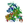
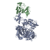
Journal: Sci Adv / Year: 2024 Title: The crystal and cryo-EM structures of PLCγ2 reveal dynamic interdomain recognitions in autoinhibition. Authors: Young-Cheul Shin / Ashlee Marie Plummer-Medeiros / Alison Mungenast / Hyeong-Wook Choi / Karen TenDyke / Xiaojie Zhu / Jennifer Shepard / Kristen Sanders / Ningning Zhuang / Liang Hu / ...Authors: Young-Cheul Shin / Ashlee Marie Plummer-Medeiros / Alison Mungenast / Hyeong-Wook Choi / Karen TenDyke / Xiaojie Zhu / Jennifer Shepard / Kristen Sanders / Ningning Zhuang / Liang Hu / Dongming Qian / Kangkang Song / Chen Xu / John Wang / Suresh B Poda / Maofu Liao / Yu Chen / Abstract: Phospholipase C gamma 2 (PLCγ2) plays important roles in cell signaling downstream of various membrane receptors. PLCγ2 contains a multidomain inhibitory region critical for its regulation, while ...Phospholipase C gamma 2 (PLCγ2) plays important roles in cell signaling downstream of various membrane receptors. PLCγ2 contains a multidomain inhibitory region critical for its regulation, while it has remained unclear how these domains contribute to PLCγ2 activity modulation. Here we determined three structures of human PLCγ2 in autoinhibited states, which reveal dynamic interactions at the autoinhibition interface, involving the conformational flexibility of the Src homology 3 (SH3) domain in the inhibitory region, and its previously unknown interaction with a carboxyl-terminal helical domain in the core region. We also determined a structure of PLCγ2 bound to the kinase domain of fibroblast growth factor receptor 1 (FGFR1), which demonstrates the recognition of FGFR1 by the nSH2 domain in the inhibitory region of PLCγ2. Our results provide structural insights into PLCγ2 regulation that will facilitate future mechanistic studies to understand the entire activation process.
Mass: 136061.469 Da / Num. of mol.: 1 Source method: isolated from a genetically manipulated source Source: (gene. exp.) Homo sapiens (human) / Gene: PLCG2 / Production host: Trichoplusia ni (cabbage looper) References: UniProt: P16885, phosphoinositide phospholipase C
In the structure databanks used in Yorodumi, some data are registered as the other names, "COVID-19 virus" and "2019-nCoV". Here are the details of the virus and the list of structure data.
Jan 31, 2019. EMDB accession codes are about to change! (news from PDBe EMDB page)
EMDB accession codes are about to change! (news from PDBe EMDB page)
The allocation of 4 digits for EMDB accession codes will soon come to an end. Whilst these codes will remain in use, new EMDB accession codes will include an additional digit and will expand incrementally as the available range of codes is exhausted. The current 4-digit format prefixed with “EMD-” (i.e. EMD-XXXX) will advance to a 5-digit format (i.e. EMD-XXXXX), and so on. It is currently estimated that the 4-digit codes will be depleted around Spring 2019, at which point the 5-digit format will come into force.
The EM Navigator/Yorodumi systems omit the EMD- prefix.
Related info.:Q: What is EMD? / ID/Accession-code notation in Yorodumi/EM Navigator
Yorodumi is a browser for structure data from EMDB, PDB, SASBDB, etc.
This page is also the successor to EM Navigator detail page, and also detail information page/front-end page for Omokage search.
The word "yorodu" (or yorozu) is an old Japanese word meaning "ten thousand". "mi" (miru) is to see.
Related info.:EMDB / PDB / SASBDB / Comparison of 3 databanks / Yorodumi Search / Aug 31, 2016. New EM Navigator & Yorodumi / Yorodumi Papers / Jmol/JSmol / Function and homology information / Changes in new EM Navigator and Yorodumi
 Movie
Movie Controller


 Open data
 Basic information
 Components
 Keywords
 Function and homology information
 Homo sapiens (human)
 X-RAY DIFFRACTION /
  Authors
 Citation

 Structure visualization
 Downloads & links
 Other downloads


 PDBj
PDBj


 Assembly
 Trichoplusia ni (cabbage looper)
 Mass: 40.078 Da / Num. of mol.: 1 / Source method: obtained synthetically / Formula: Ca / Feature type: SUBJECT OF INVESTIGATION
 Mass: 62.068 Da / Num. of mol.: 1 / Source method: obtained synthetically / Formula: C2H6O2
 Mass: 18.015 Da / Num. of mol.: 190 / Source method: isolated from a natural source / Formula: H2O
 Sample preparation
 Processing