 Movie
Movie Controller


+ Open data

- Basic information
| Entry | Database: PDB / ID: 8sik | |||||||||||||||||||||||||||||||||||||||
|---|---|---|---|---|---|---|---|---|---|---|---|---|---|---|---|---|---|---|---|---|---|---|---|---|---|---|---|---|---|---|---|---|---|---|---|---|---|---|---|---|
| Title | KCNQ1 with voltage sensor in the up conformation | |||||||||||||||||||||||||||||||||||||||
 Components Components |
| |||||||||||||||||||||||||||||||||||||||
 Keywords Keywords | MEMBRANE PROTEIN / voltage-gated potassium channel | |||||||||||||||||||||||||||||||||||||||
| Function / homology |  Function and homology information Function and homology informationgastrin-induced gastric acid secretion / corticosterone secretion / voltage-gated potassium channel activity involved in atrial cardiac muscle cell action potential repolarization / negative regulation of voltage-gated potassium channel activity / basolateral part of cell / lumenal side of membrane / negative regulation of delayed rectifier potassium channel activity / rhythmic behavior / stomach development / regulation of gastric acid secretion ...gastrin-induced gastric acid secretion / corticosterone secretion / voltage-gated potassium channel activity involved in atrial cardiac muscle cell action potential repolarization / negative regulation of voltage-gated potassium channel activity / basolateral part of cell / lumenal side of membrane / negative regulation of delayed rectifier potassium channel activity / rhythmic behavior / stomach development / regulation of gastric acid secretion / voltage-gated potassium channel activity involved in cardiac muscle cell action potential repolarization / Phase 3 - rapid repolarisation / membrane repolarization during action potential / membrane repolarization during atrial cardiac muscle cell action potential / Phase 2 - plateau phase / iodide transport / regulation of atrial cardiac muscle cell membrane repolarization / membrane repolarization during ventricular cardiac muscle cell action potential / intracellular chloride ion homeostasis / potassium ion export across plasma membrane / membrane repolarization during cardiac muscle cell action potential / voltage-gated potassium channel activity involved in ventricular cardiac muscle cell action potential repolarization / atrial cardiac muscle cell action potential / regulation of membrane repolarization / renal sodium ion absorption / auditory receptor cell development / protein phosphatase 1 binding / detection of mechanical stimulus involved in sensory perception of sound / delayed rectifier potassium channel activity / ventricular cardiac muscle cell action potential / potassium ion homeostasis / regulation of ventricular cardiac muscle cell membrane repolarization / positive regulation of potassium ion transmembrane transport / Voltage gated Potassium channels / non-motile cilium assembly / outward rectifier potassium channel activity / intestinal absorption / CaM pathway / Cam-PDE 1 activation / Sodium/Calcium exchangers / Calmodulin induced events / cardiac muscle cell contraction / inner ear morphogenesis / Reduction of cytosolic Ca++ levels / Activation of Ca-permeable Kainate Receptor / CREB1 phosphorylation through the activation of CaMKII/CaMKK/CaMKIV cascasde / Loss of phosphorylation of MECP2 at T308 / CREB1 phosphorylation through the activation of Adenylate Cyclase / adrenergic receptor signaling pathway / negative regulation of high voltage-gated calcium channel activity / PKA activation / CaMK IV-mediated phosphorylation of CREB / Glycogen breakdown (glycogenolysis) / CLEC7A (Dectin-1) induces NFAT activation / negative regulation of ryanodine-sensitive calcium-release channel activity / organelle localization by membrane tethering / Activation of RAC1 downstream of NMDARs / renal absorption / : / autophagosome membrane docking / ciliary base / protein kinase A regulatory subunit binding / negative regulation of calcium ion export across plasma membrane / regulation of ryanodine-sensitive calcium-release channel activity / regulation of cardiac muscle cell action potential / protein kinase A catalytic subunit binding / presynaptic endocytosis / regulation of heart contraction / inner ear development / Synthesis of IP3 and IP4 in the cytosol / Phase 0 - rapid depolarisation / potassium ion import across plasma membrane / Negative regulation of NMDA receptor-mediated neuronal transmission / regulation of heart rate by cardiac conduction / Unblocking of NMDA receptors, glutamate binding and activation / RHO GTPases activate PAKs / calcineurin-mediated signaling / regulation of cell communication by electrical coupling involved in cardiac conduction / monoatomic ion channel complex / Ion transport by P-type ATPases / Uptake and function of anthrax toxins / protein phosphatase activator activity / cochlea development / Long-term potentiation / Calcineurin activates NFAT / action potential / Regulation of MECP2 expression and activity / DARPP-32 events / Smooth Muscle Contraction / voltage-gated potassium channel activity / detection of calcium ion / social behavior / regulation of cardiac muscle contraction / positive regulation of heart rate / catalytic complex / RHO GTPases activate IQGAPs / transport vesicle / calcium channel inhibitor activity / presynaptic cytosol / Activation of AMPK downstream of NMDARs Similarity search - Function | |||||||||||||||||||||||||||||||||||||||
| Biological species |  Homo sapiens (human) Homo sapiens (human) | |||||||||||||||||||||||||||||||||||||||
| Method | ELECTRON MICROSCOPY / single particle reconstruction / cryo EM / Resolution: 2.9 Å | |||||||||||||||||||||||||||||||||||||||
 Authors Authors | Mandala, V.S. / MacKinnon, R. | |||||||||||||||||||||||||||||||||||||||
| Funding support |  United States, 1items United States, 1items
| |||||||||||||||||||||||||||||||||||||||
 Citation Citation | Journal: Proc Natl Acad Sci U S A / Year: 2023 Title: The membrane electric field regulates the PIP-binding site to gate the KCNQ1 channel. Authors: Venkata Shiva Mandala / Roderick MacKinnon / Abstract: Voltage-dependent ion channels underlie the propagation of action potentials and other forms of electrical activity in cells. In these proteins, voltage sensor domains (VSDs) regulate opening and ...Voltage-dependent ion channels underlie the propagation of action potentials and other forms of electrical activity in cells. In these proteins, voltage sensor domains (VSDs) regulate opening and closing of the pore through the displacement of their positive-charged S4 helix in response to the membrane voltage. The movement of S4 at hyperpolarizing membrane voltages in some channels is thought to directly clamp the pore shut through the S4-S5 linker helix. The KCNQ1 channel (also known as K7.1), which is important for heart rhythm, is regulated not only by membrane voltage but also by the signaling lipid phosphatidylinositol 4,5-bisphosphate (PIP). KCNQ1 requires PIP to open and to couple the movement of S4 in the VSD to the pore. To understand the mechanism of this voltage regulation, we use cryogenic electron microscopy to visualize the movement of S4 in the human KCNQ1 channel in lipid membrane vesicles with a voltage difference across the membrane, i.e., an applied electric field in the membrane. Hyperpolarizing voltages displace S4 in such a manner as to sterically occlude the PIP-binding site. Thus, in KCNQ1, the voltage sensor acts primarily as a regulator of PIP binding. The voltage sensors' influence on the channel's gate is indirect through the reaction sequence: voltage sensor movement → alter PIP ligand affinity → alter pore opening. | |||||||||||||||||||||||||||||||||||||||
| History |
|
- Structure visualization
| Structure viewer | Molecule: MolmilJmol/JSmol |
|---|
- Downloads & links
-Download
| PDBx/mmCIF format | 8sik.cif.gz | 400.3 KB | Display | PDBx/mmCIF format |
|---|---|---|---|---|
| PDB format | pdb8sik.ent.gz | 320.3 KB | Display | PDB format |
| PDBx/mmJSON format | 8sik.json.gz | Tree view | PDBx/mmJSON format | |
| Others |  Other downloads Other downloads |
-Validation report
| Arichive directory | https://data.pdbj.org/pub/pdb/validation_reports/si/8sikftp://data.pdbj.org/pub/pdb/validation_reports/si/8sik | HTTPS FTP |
|---|
-Related structure data
| Related structure data |  40508MC 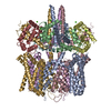 8simC  8sinC M: map data used to model this data C: citing same article ( |
|---|---|
| Similar structure data |
-Links
 PDBj
PDBj


- Assembly
| Deposited unit | 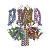
|
|---|---|
| 1 |
|
-Components
| #1: Protein | Mass: 16852.545 Da / Num. of mol.: 4 Source method: isolated from a genetically manipulated source Source: (gene. exp.) Homo sapiens (human) / Gene: CALM1, CALM, CAM, CAM1 / Production host: Homo sapiens (human) / References: UniProt: P0DP23#2: Protein | Mass: 63258.574 Da / Num. of mol.: 4 Source method: isolated from a genetically manipulated source Source: (gene. exp.) Homo sapiens (human) / Gene: KCNQ1, KCNA8, KCNA9, KVLQT1 / Production host: Homo sapiens (human) / References: UniProt: P51787#3: Chemical | ChemComp-CA /   Mass: 40.078 Da / Num. of mol.: 8 / Source method: obtained synthetically / Formula: Ca Mass: 40.078 Da / Num. of mol.: 8 / Source method: obtained synthetically / Formula: CaHas ligand of interest | N | Has protein modification | N | |
|---|
-Experimental details
-Experiment
| Experiment | Method: ELECTRON MICROSCOPY |
|---|---|
| EM experiment | Aggregation state: PARTICLE / 3D reconstruction method: single particle reconstruction |
- Sample preparation
| Component | Name: Complex of KCNQ1 (Kv7.1) channel bound to calmodulin-Ca2+ Type: COMPLEX / Entity ID: #1-#2 / Source: RECOMBINANT |
|---|---|
| Molecular weight | Experimental value: NO |
| Source (natural) | Organism: Homo sapiens (human) |
| Source (recombinant) | Organism: Homo sapiens (human) |
| Buffer solution | pH: 8 |
| Specimen | Conc.: 0.2 mg/ml / Embedding applied: NO / Shadowing applied: NO / Staining applied: NO / Vitrification applied: YES |
| Vitrification | Instrument: FEI VITROBOT MARK IV / Cryogen name: ETHANE / Humidity: 100 % / Chamber temperature: 293 K |
- Electron microscopy imaging
| Experimental equipment |  Model: Titan Krios / Image courtesy: FEI Company |
|---|---|
| Microscopy | Model: FEI TITAN KRIOS |
| Electron gun | Electron source:  FIELD EMISSION GUN / Accelerating voltage: 300 kV / Illumination mode: FLOOD BEAM FIELD EMISSION GUN / Accelerating voltage: 300 kV / Illumination mode: FLOOD BEAM |
| Electron lens | Mode: BRIGHT FIELD / Nominal defocus max: 2000 nm / Nominal defocus min: 1000 nm |
| Image recording | Electron dose: 60 e/Å2 / Film or detector model: GATAN K3 BIOQUANTUM (6k x 4k) |
- Processing
| Software | Name: PHENIX / Version: 1.20.1_4487: / Classification: refinement | ||||||||||||||||||||||||
|---|---|---|---|---|---|---|---|---|---|---|---|---|---|---|---|---|---|---|---|---|---|---|---|---|---|
| EM software | Name: PHENIX / Category: model refinement | ||||||||||||||||||||||||
| CTF correction | Type: PHASE FLIPPING AND AMPLITUDE CORRECTION | ||||||||||||||||||||||||
| Symmetry | Point symmetry: C4 (4 fold cyclic) | ||||||||||||||||||||||||
| 3D reconstruction | Resolution: 2.9 Å / Resolution method: FSC 0.143 CUT-OFF / Num. of particles: 200862 / Symmetry type: POINT | ||||||||||||||||||||||||
| Refine LS restraints |
|
