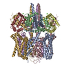
Journal: Proc Natl Acad Sci U S A / Year: 2023 Title: The membrane electric field regulates the PIP-binding site to gate the KCNQ1 channel. Authors: Venkata Shiva Mandala / Roderick MacKinnon / Abstract: Voltage-dependent ion channels underlie the propagation of action potentials and other forms of electrical activity in cells. In these proteins, voltage sensor domains (VSDs) regulate opening and ...Voltage-dependent ion channels underlie the propagation of action potentials and other forms of electrical activity in cells. In these proteins, voltage sensor domains (VSDs) regulate opening and closing of the pore through the displacement of their positive-charged S4 helix in response to the membrane voltage. The movement of S4 at hyperpolarizing membrane voltages in some channels is thought to directly clamp the pore shut through the S4-S5 linker helix. The KCNQ1 channel (also known as K7.1), which is important for heart rhythm, is regulated not only by membrane voltage but also by the signaling lipid phosphatidylinositol 4,5-bisphosphate (PIP). KCNQ1 requires PIP to open and to couple the movement of S4 in the VSD to the pore. To understand the mechanism of this voltage regulation, we use cryogenic electron microscopy to visualize the movement of S4 in the human KCNQ1 channel in lipid membrane vesicles with a voltage difference across the membrane, i.e., an applied electric field in the membrane. Hyperpolarizing voltages displace S4 in such a manner as to sterically occlude the PIP-binding site. Thus, in KCNQ1, the voltage sensor acts primarily as a regulator of PIP binding. The voltage sensors' influence on the channel's gate is indirect through the reaction sequence: voltage sensor movement → alter PIP ligand affinity → alter pore opening.
In the structure databanks used in Yorodumi, some data are registered as the other names, "COVID-19 virus" and "2019-nCoV". Here are the details of the virus and the list of structure data.
Jan 31, 2019. EMDB accession codes are about to change! (news from PDBe EMDB page)
EMDB accession codes are about to change! (news from PDBe EMDB page)
The allocation of 4 digits for EMDB accession codes will soon come to an end. Whilst these codes will remain in use, new EMDB accession codes will include an additional digit and will expand incrementally as the available range of codes is exhausted. The current 4-digit format prefixed with “EMD-” (i.e. EMD-XXXX) will advance to a 5-digit format (i.e. EMD-XXXXX), and so on. It is currently estimated that the 4-digit codes will be depleted around Spring 2019, at which point the 5-digit format will come into force.
The EM Navigator/Yorodumi systems omit the EMD- prefix.
Related info.:Q: What is EMD? / ID/Accession-code notation in Yorodumi/EM Navigator
Yorodumi is a browser for structure data from EMDB, PDB, SASBDB, etc.
This page is also the successor to EM Navigator detail page, and also detail information page/front-end page for Omokage search.
The word "yorodu" (or yorozu) is an old Japanese word meaning "ten thousand". "mi" (miru) is to see.
Related info.:EMDB / PDB / SASBDB / Comparison of 3 databanks / Yorodumi Search / Aug 31, 2016. New EM Navigator & Yorodumi / Yorodumi Papers / Jmol/JSmol / Function and homology information / Changes in new EM Navigator and Yorodumi
 Movie
Movie Controller


 Open data
 Basic information
 Map data
 Sample
 Keywords
 Function and homology information
 Homo sapiens (human)
 Authors
 United States, 1 items
  Citation
 Structure visualization
 Downloads & links
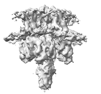 emd_40510.png
 http://ftp.pdbj.org/pub/emdb/structures/EMD-40510


 Z (Sec.)
 Y (Row.)
 X (Col.)


 Sample components
 Processing
 Electron microscopy
 FIELD EMISSION GUN
