 Movie
Movie Controller

[English] 日本語
 Yorodumi
- PDB-8s8a: Human pyridoxal phosphatase in complex with 7,8-dihydroxyflavone ... -
+ Open data

- Basic information
| Entry | Database: PDB / ID: 8s8a | ||||||
|---|---|---|---|---|---|---|---|
| Title | Human pyridoxal phosphatase in complex with 7,8-dihydroxyflavone without phosphate | ||||||
 Components Components | Chronophin | ||||||
 Keywords Keywords | HYDROLASE / Inhibitor / PDXP / 7 8-dihydroxyflavone / Flavone / Chronophin | ||||||
| Function / homology |  Function and homology information Function and homology informationpyridoxal phosphatase / pyridoxal phosphate catabolic process / actin rod assembly / pyridoxal phosphatase activity / positive regulation of actin filament depolymerization / regulation of modification of postsynaptic structure / dephosphorylation / regulation of mitotic nuclear division / protein dephosphorylation / protein-serine/threonine phosphatase ...pyridoxal phosphatase / pyridoxal phosphate catabolic process / actin rod assembly / pyridoxal phosphatase activity / positive regulation of actin filament depolymerization / regulation of modification of postsynaptic structure / dephosphorylation / regulation of mitotic nuclear division / protein dephosphorylation / protein-serine/threonine phosphatase / cellular response to ATP / protein serine/threonine phosphatase activity / lamellipodium membrane / heat shock protein binding / phosphoprotein phosphatase activity / regulation of cytokinesis / ruffle membrane / cell-cell junction / cytoskeleton / postsynapse / glutamatergic synapse / magnesium ion binding / protein homodimerization activity / cytoplasm / cytosol Similarity search - Function | ||||||
| Biological species |  Homo sapiens (human) Homo sapiens (human) | ||||||
| Method |  X-RAY DIFFRACTION / SYNCHROTRON / MOLECULAR REPLACEMENT / Resolution: 1.5 Å X-RAY DIFFRACTION / SYNCHROTRON / MOLECULAR REPLACEMENT / Resolution: 1.5 Å | ||||||
 Authors Authors | Brenner, M. / Gohla, A. / Schindelin, H. | ||||||
| Funding support | 1items
| ||||||
 Citation Citation | Journal: Elife / Year: 2024 Title: 7,8-Dihydroxyflavone is a direct inhibitor of human and murine pyridoxal phosphatase. Authors: Brenner, M. / Zink, C. / Witzinger, L. / Keller, A. / Hadamek, K. / Bothe, S. / Neuenschwander, M. / Villmann, C. / von Kries, J.P. / Schindelin, H. / Jeanclos, E. / Gohla, A. #1: Journal: Elife / Year: 2024Title: 7,8-Dihydroxyflavone is a direct inhibitor of pyridoxal phosphatase Authors: Brenner, M. / Zink, C. / Witzinger, L. / Keller, A. / Hadamek, K. / Bothe, S. / Neuenschwander, M. / Villmann, C. / von Kries, J.P. / Schindelin, H. / Jeanclos, E. / Gohla, A. #2: Journal: Acta Crystallogr.,Sect.D / Year: 2012 Title: Towards automated crystallographic structure refinement with phenix.refine. Authors: Afonine, P.V. / Grosse-Kunstleve, R.W. / Echols, N. / Headd, J.J. / Moriarty, N.W. / Mustyakimov, M. / Terwilliger, T.C. / Urzhumtsev, A. / Zwart, P.H. / Adams, P.D. #3: Journal: Acta Crystallogr D Struct Biol / Year: 2019 Title: Macromolecular structure determination using X-rays, neutrons and electrons: recent developments in Phenix. Authors: Dorothee Liebschner / Pavel V Afonine / Matthew L Baker / Gábor Bunkóczi / Vincent B Chen / Tristan I Croll / Bradley Hintze / Li Wei Hung / Swati Jain / Airlie J McCoy / Nigel W Moriarty ...Authors: Dorothee Liebschner / Pavel V Afonine / Matthew L Baker / Gábor Bunkóczi / Vincent B Chen / Tristan I Croll / Bradley Hintze / Li Wei Hung / Swati Jain / Airlie J McCoy / Nigel W Moriarty / Robert D Oeffner / Billy K Poon / Michael G Prisant / Randy J Read / Jane S Richardson / David C Richardson / Massimo D Sammito / Oleg V Sobolev / Duncan H Stockwell / Thomas C Terwilliger / Alexandre G Urzhumtsev / Lizbeth L Videau / Christopher J Williams / Paul D Adams /    Abstract: Diffraction (X-ray, neutron and electron) and electron cryo-microscopy are powerful methods to determine three-dimensional macromolecular structures, which are required to understand biological ...Diffraction (X-ray, neutron and electron) and electron cryo-microscopy are powerful methods to determine three-dimensional macromolecular structures, which are required to understand biological processes and to develop new therapeutics against diseases. The overall structure-solution workflow is similar for these techniques, but nuances exist because the properties of the reduced experimental data are different. Software tools for structure determination should therefore be tailored for each method. Phenix is a comprehensive software package for macromolecular structure determination that handles data from any of these techniques. Tasks performed with Phenix include data-quality assessment, map improvement, model building, the validation/rebuilding/refinement cycle and deposition. Each tool caters to the type of experimental data. The design of Phenix emphasizes the automation of procedures, where possible, to minimize repetitive and time-consuming manual tasks, while default parameters are chosen to encourage best practice. A graphical user interface provides access to many command-line features of Phenix and streamlines the transition between programs, project tracking and re-running of previous tasks. | ||||||
| History |
|
- Structure visualization
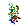
| Structure viewer | Molecule: MolmilJmol/JSmol |
|---|
- Downloads & links
-Download
| PDBx/mmCIF format | 8s8a.cif.gz | 237 KB | Display | PDBx/mmCIF format |
|---|---|---|---|---|
| PDB format | pdb8s8a.ent.gz | 160.1 KB | Display | PDB format |
| PDBx/mmJSON format | 8s8a.json.gz | Tree view | PDBx/mmJSON format | |
| Others |  Other downloads Other downloads |
-Validation report
| Arichive directory | https://data.pdbj.org/pub/pdb/validation_reports/s8/8s8aftp://data.pdbj.org/pub/pdb/validation_reports/s8/8s8a | HTTPS FTP |
|---|
-Related structure data

-Links
 PDBj
PDBj- Assembly
| Deposited unit | 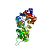
| ||||||||||||
|---|---|---|---|---|---|---|---|---|---|---|---|---|---|
| 1 | 
| ||||||||||||
| Unit cell |
| ||||||||||||
| Components on special symmetry positions |
|
-Components
-Protein , 1 types, 1 molecules A
| #1: Protein | Mass: 31738.145 Da / Num. of mol.: 1 Source method: isolated from a genetically manipulated source Source: (gene. exp.) Homo sapiens (human) / Gene: PDXP, CIN, PLP, PLPP / Production host:  References: UniProt: Q96GD0, protein-serine/threonine phosphatase, pyridoxal phosphatase |
|---|
-Non-polymers , 5 types, 274 molecules 


| #2: Chemical | ChemComp-UK9 / Mass: 254.237 Da / Num. of mol.: 1 / Source method: obtained synthetically / Formula: C15H10O4 / Feature type: SUBJECT OF INVESTIGATION |
|---|---|
| #3: Chemical | ChemComp-FMT /  Mass: 46.025 Da / Num. of mol.: 1 / Source method: obtained synthetically / Formula: CH2O2 Mass: 46.025 Da / Num. of mol.: 1 / Source method: obtained synthetically / Formula: CH2O2 |
| #4: Chemical | ChemComp-MG /  Mass: 24.305 Da / Num. of mol.: 1 / Source method: obtained synthetically / Formula: Mg Mass: 24.305 Da / Num. of mol.: 1 / Source method: obtained synthetically / Formula: Mg |
| #5: Chemical | ChemComp-CL /  Mass: 35.453 Da / Num. of mol.: 1 / Source method: obtained synthetically / Formula: Cl Mass: 35.453 Da / Num. of mol.: 1 / Source method: obtained synthetically / Formula: Cl |
| #6: Water | ChemComp-HOH / Mass: 18.015 Da / Num. of mol.: 270 / Source method: isolated from a natural source / Formula: H2O |
-Details
| Has ligand of interest | Y |
|---|---|
| Has protein modification | Y |
-Experimental details
-Experiment
| Experiment | Method: X-RAY DIFFRACTION / Number of used crystals: 1 |
|---|
- Sample preparation
| Crystal | Density Matthews: 2.32 Å3/Da / Density % sol: 47 % |
|---|---|
| Crystal grow | Temperature: 293 K / Method: vapor diffusion, sitting drop / pH: 7 Details: PDXP at 10 mg/ml in 50 mM Triethanolamine, 250 mM NaCl and 5 mM MgCl2 was crystallized against 0.1 M HEPES, 15 % Tacsimate, 2 % PEG 3350 |
-Data collection
| Diffraction | Mean temperature: 100 K / Serial crystal experiment: N |
|---|---|
| Diffraction source | Source: SYNCHROTRON / Site: ESRF / Beamline: ID23-2 / Wavelength: 0.873128 Å |
| Detector | Type: DECTRIS EIGER X 9M / Detector: PIXEL / Date: Feb 16, 2024 |
| Radiation | Monochromator: Si(111) / Protocol: SINGLE WAVELENGTH / Monochromatic (M) / Laue (L): M / Scattering type: x-ray |
| Radiation wavelength | Wavelength: 0.873128 Å / Relative weight: 1 |
| Reflection | Resolution: 1.5→48.17 Å / Num. obs: 51834 / % possible obs: 100 % / Redundancy: 25.6 % / Biso Wilson estimate: 28.34 Å2 / CC1/2: 0.999 / Rmerge(I) obs: 0.081 / Rpim(I) all: 0.017 / Rrim(I) all: 0.083 / Net I/σ(I): 17.5 |
| Reflection shell | Resolution: 1.5→1.53 Å / Redundancy: 26 % / Rmerge(I) obs: 3.67 / Mean I/σ(I) obs: 1.1 / Num. unique obs: 2480 / CC1/2: 0.579 / Rpim(I) all: 0.731 / Rrim(I) all: 3.743 / % possible all: 100 |
- Processing
| Software |
| |||||||||||||||||||||||||||||||||||||||||||||||||||||||||||||||||||||||||||||||||||||||||||||||||||||||||||||||||||||||||||||||||||||
|---|---|---|---|---|---|---|---|---|---|---|---|---|---|---|---|---|---|---|---|---|---|---|---|---|---|---|---|---|---|---|---|---|---|---|---|---|---|---|---|---|---|---|---|---|---|---|---|---|---|---|---|---|---|---|---|---|---|---|---|---|---|---|---|---|---|---|---|---|---|---|---|---|---|---|---|---|---|---|---|---|---|---|---|---|---|---|---|---|---|---|---|---|---|---|---|---|---|---|---|---|---|---|---|---|---|---|---|---|---|---|---|---|---|---|---|---|---|---|---|---|---|---|---|---|---|---|---|---|---|---|---|---|---|---|
| Refinement | Method to determine structure: MOLECULAR REPLACEMENT / Resolution: 1.5→48.17 Å / SU ML: 0.1944 / Cross valid method: FREE R-VALUE / σ(F): 1.34 / Phase error: 21.2262 Stereochemistry target values: GeoStd + Monomer Library + CDL v1.2
| |||||||||||||||||||||||||||||||||||||||||||||||||||||||||||||||||||||||||||||||||||||||||||||||||||||||||||||||||||||||||||||||||||||
| Solvent computation | Shrinkage radii: 0.9 Å / VDW probe radii: 1.1 Å / Solvent model: FLAT BULK SOLVENT MODEL | |||||||||||||||||||||||||||||||||||||||||||||||||||||||||||||||||||||||||||||||||||||||||||||||||||||||||||||||||||||||||||||||||||||
| Displacement parameters | Biso mean: 37.21 Å2 | |||||||||||||||||||||||||||||||||||||||||||||||||||||||||||||||||||||||||||||||||||||||||||||||||||||||||||||||||||||||||||||||||||||
| Refinement step | Cycle: LAST / Resolution: 1.5→48.17 Å
| |||||||||||||||||||||||||||||||||||||||||||||||||||||||||||||||||||||||||||||||||||||||||||||||||||||||||||||||||||||||||||||||||||||
| Refine LS restraints |
| |||||||||||||||||||||||||||||||||||||||||||||||||||||||||||||||||||||||||||||||||||||||||||||||||||||||||||||||||||||||||||||||||||||
| LS refinement shell |
| |||||||||||||||||||||||||||||||||||||||||||||||||||||||||||||||||||||||||||||||||||||||||||||||||||||||||||||||||||||||||||||||||||||
| Refinement TLS params. | Method: refined / Refine-ID: X-RAY DIFFRACTION
| |||||||||||||||||||||||||||||||||||||||||||||||||||||||||||||||||||||||||||||||||||||||||||||||||||||||||||||||||||||||||||||||||||||
| Refinement TLS group | Refine-ID: X-RAY DIFFRACTION / Auth asym-ID: A / Label asym-ID: A
|
