Movie
Movie Controller


+ Open data

- Basic information
| Entry | Database: PDB / ID: 8s0e | |||||||||||||||
|---|---|---|---|---|---|---|---|---|---|---|---|---|---|---|---|---|
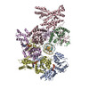
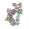
| Title | H. sapiens OCCM bound to double stranded DNA | |||||||||||||||
 Components Components |
| |||||||||||||||
 Keywords Keywords | REPLICATION / AAA+ ATPase / DNA helicase | |||||||||||||||
| Function / homology |  Function and homology information Function and homology informationDNA replication preinitiation complex assembly / mitotic pre-replicative complex assembly / cellular response to vasopressin / response to sorbitol / positive regulation of chromosome segregation / CDC6 association with the ORC:origin complex / polar body extrusion after meiotic divisions / origin recognition complex / regulation of nuclear cell cycle DNA replication / E2F-enabled inhibition of pre-replication complex formation ...DNA replication preinitiation complex assembly / mitotic pre-replicative complex assembly / cellular response to vasopressin / response to sorbitol / positive regulation of chromosome segregation / CDC6 association with the ORC:origin complex / polar body extrusion after meiotic divisions / origin recognition complex / regulation of nuclear cell cycle DNA replication / E2F-enabled inhibition of pre-replication complex formation / inner kinetochore / Switching of origins to a post-replicative state / Unwinding of DNA / traversing start control point of mitotic cell cycle / nuclear origin of replication recognition complex / regulation of phosphorylation / negative regulation of DNA-templated DNA replication / alpha DNA polymerase:primase complex / Regulation of MITF-M-dependent genes involved in DNA replication, damage repair and senescence / attachment of mitotic spindle microtubules to kinetochore / nuclear pre-replicative complex / CMG complex / DNA replication checkpoint signaling / DNA replication preinitiation complex / mitotic DNA replication checkpoint signaling / regulation of cyclin-dependent protein serine/threonine kinase activity / double-strand break repair via break-induced replication / MCM complex / mitotic DNA replication initiation / Transcription of E2F targets under negative control by DREAM complex / mitotic DNA replication / regulation of mitotic metaphase/anaphase transition / regulation of DNA-templated DNA replication initiation / DNA strand elongation involved in DNA replication / G1/S-Specific Transcription / positive regulation of cytokinesis / spindle midzone / negative regulation of DNA replication / intercellular bridge / negative regulation of cell cycle / regulation of DNA replication / cellular response to angiotensin / DNA replication origin binding / cochlea development / protein polymerization / Activation of the pre-replicative complex / DNA replication initiation / Activation of ATR in response to replication stress / heterochromatin / DNA polymerase binding / cellular response to epidermal growth factor stimulus / positive regulation of DNA replication / DNA helicase activity / Assembly of the ORC complex at the origin of replication / protein serine/threonine kinase binding / Assembly of the pre-replicative complex / positive regulation of fibroblast proliferation / CDK-mediated phosphorylation and removal of Cdc6 / mitotic spindle / kinetochore / spindle pole / Orc1 removal from chromatin / mitotic cell cycle / single-stranded DNA binding / histone binding / DNA helicase / chromosome, telomeric region / DNA replication / nuclear body / response to xenobiotic stimulus / negative regulation of cell population proliferation / cell division / nucleotide binding / centrosome / chromatin binding / apoptotic process / DNA damage response / nucleolus / chromatin / perinuclear region of cytoplasm / negative regulation of transcription by RNA polymerase II / enzyme binding / ATP hydrolysis activity / DNA binding / nucleoplasm / zinc ion binding / ATP binding / membrane / metal ion binding / identical protein binding / nucleus / cytosol / cytoplasm Similarity search - Function | |||||||||||||||
| Biological species |  Homo sapiens (human) Homo sapiens (human)synthetic construct (others) | |||||||||||||||
| Method | ELECTRON MICROSCOPY / single particle reconstruction / cryo EM / Resolution: 3.8 Å | |||||||||||||||
 Authors Authors | Greiwe, J.F. / Weissmann, F. / Diffley, J.F.X. / Costa, A. | |||||||||||||||
| Funding support |  United Kingdom, European Union, 4items United Kingdom, European Union, 4items
| |||||||||||||||
 Citation Citation | Journal: Nature / Year: 2024 Title: MCM double hexamer loading visualized with human proteins. Authors: Florian Weissmann / Julia F Greiwe / Thomas Pühringer / Evelyn L Eastwood / Emma C Couves / Thomas C R Miller / John F X Diffley / Alessandro Costa /  Abstract: Eukaryotic DNA replication begins with the loading of the MCM replicative DNA helicase as a head-to-head double hexamer at origins of DNA replication. Our current understanding of how the double ...Eukaryotic DNA replication begins with the loading of the MCM replicative DNA helicase as a head-to-head double hexamer at origins of DNA replication. Our current understanding of how the double hexamer is assembled by the origin recognition complex (ORC), CDC6 and CDT1 comes mostly from budding yeast. Here we characterize human double hexamer (hDH) loading using biochemical reconstitution and cryo-electron microscopy with purified proteins. We show that the human double hexamer engages DNA differently from the yeast double hexamer (yDH), and generates approximately five base pairs of underwound DNA at the interface between hexamers, as seen in hDH isolated from cells. We identify several differences from the yeast double hexamer in the order of factor recruitment and dependencies during hDH assembly. Unlike in yeast, the ORC6 subunit of the ORC is not essential for initial MCM recruitment or hDH loading, but contributes to an alternative hDH assembly pathway that requires an intrinsically disordered region in ORC1, which may work through a MCM-ORC intermediate. Our work presents a detailed view of how double hexamers are assembled in an organism that uses sequence-independent replication origins, provides further evidence for diversity in eukaryotic double hexamer assembly mechanisms, and represents a first step towards reconstitution of DNA replication initiation with purified human proteins. #1: Journal: Biorxiv / Year: 2024Title: MCM Double Hexamer Loading Visualised with Human Proteins Authors: Weissmann, F. / Greiwe, J.F. / Puhringer, T. / Miller, T.C.R. / Diffley, J.F.X. / Costa, A. #2: Journal: Acta Crystallogr D Struct Biol / Year: 2019 Title: Macromolecular structure determination using X-rays, neutrons and electrons: recent developments in Phenix. Authors: Dorothee Liebschner / Pavel V Afonine / Matthew L Baker / Gábor Bunkóczi / Vincent B Chen / Tristan I Croll / Bradley Hintze / Li Wei Hung / Swati Jain / Airlie J McCoy / Nigel W Moriarty ...Authors: Dorothee Liebschner / Pavel V Afonine / Matthew L Baker / Gábor Bunkóczi / Vincent B Chen / Tristan I Croll / Bradley Hintze / Li Wei Hung / Swati Jain / Airlie J McCoy / Nigel W Moriarty / Robert D Oeffner / Billy K Poon / Michael G Prisant / Randy J Read / Jane S Richardson / David C Richardson / Massimo D Sammito / Oleg V Sobolev / Duncan H Stockwell / Thomas C Terwilliger / Alexandre G Urzhumtsev / Lizbeth L Videau / Christopher J Williams / Paul D Adams /   Abstract: Diffraction (X-ray, neutron and electron) and electron cryo-microscopy are powerful methods to determine three-dimensional macromolecular structures, which are required to understand biological ...Diffraction (X-ray, neutron and electron) and electron cryo-microscopy are powerful methods to determine three-dimensional macromolecular structures, which are required to understand biological processes and to develop new therapeutics against diseases. The overall structure-solution workflow is similar for these techniques, but nuances exist because the properties of the reduced experimental data are different. Software tools for structure determination should therefore be tailored for each method. Phenix is a comprehensive software package for macromolecular structure determination that handles data from any of these techniques. Tasks performed with Phenix include data-quality assessment, map improvement, model building, the validation/rebuilding/refinement cycle and deposition. Each tool caters to the type of experimental data. The design of Phenix emphasizes the automation of procedures, where possible, to minimize repetitive and time-consuming manual tasks, while default parameters are chosen to encourage best practice. A graphical user interface provides access to many command-line features of Phenix and streamlines the transition between programs, project tracking and re-running of previous tasks. | |||||||||||||||
| History |
|
- Structure visualization
| Structure viewer | Molecule: MolmilJmol/JSmol |
|---|
- Downloads & links
-Download
| PDBx/mmCIF format | 8s0e.cif.gz | 1.2 MB | Display | PDBx/mmCIF format |
|---|---|---|---|---|
| PDB format | pdb8s0e.ent.gz | 822.7 KB | Display | PDB format |
| PDBx/mmJSON format | 8s0e.json.gz | Tree view | PDBx/mmJSON format | |
| Others |  Other downloads Other downloads |
-Validation report
| Arichive directory | https://data.pdbj.org/pub/pdb/validation_reports/s0/8s0eftp://data.pdbj.org/pub/pdb/validation_reports/s0/8s0e | HTTPS FTP |
|---|
-Related structure data
| Related structure data |  19623MC  8s09C  8s0aC  8s0bC  8s0cC  8s0dC  8s0fC C: citing same article ( M: map data used to model this data |
|---|---|
| Similar structure data |


-Links
 PDBj
PDBj


- Assembly
| Deposited unit | 
|
|---|---|
| 1 |
|
-Components
-DNA chain , 2 types, 2 molecules XY
| #1: DNA chain | Mass: 12000.695 Da / Num. of mol.: 1 / Source method: obtained synthetically / Source: (synth.) synthetic construct (others) |
|---|---|
| #2: DNA chain | Mass: 12009.710 Da / Num. of mol.: 1 / Source method: obtained synthetically / Source: (synth.) synthetic construct (others) |
-DNA replication licensing factor ... , 6 types, 6 molecules 524673
| #3: Protein | Mass: 82406.633 Da / Num. of mol.: 1 Source method: isolated from a genetically manipulated source Source: (gene. exp.) Homo sapiens (human) / Gene: MCM5, CDC46 / Production host:   Spodoptera frugiperda (fall armyworm) / References: UniProt: P33992, DNA helicase Spodoptera frugiperda (fall armyworm) / References: UniProt: P33992, DNA helicase |
|---|---|
| #5: Protein | Mass: 102034.102 Da / Num. of mol.: 1 Source method: isolated from a genetically manipulated source Source: (gene. exp.) Homo sapiens (human) / Gene: MCM2, BM28, CCNL1, CDCL1, KIAA0030 / Production host: Spodoptera frugiperda (fall armyworm) / References: UniProt: P49736, DNA helicase |
| #6: Protein | Mass: 96702.891 Da / Num. of mol.: 1 Source method: isolated from a genetically manipulated source Source: (gene. exp.) Homo sapiens (human) / Gene: MCM4, CDC21 / Production host: Spodoptera frugiperda (fall armyworm) / References: UniProt: P33991, DNA helicase |
| #7: Protein | Mass: 93010.273 Da / Num. of mol.: 1 Source method: isolated from a genetically manipulated source Source: (gene. exp.) Homo sapiens (human) / Gene: MCM6 / Production host: Spodoptera frugiperda (fall armyworm) / References: UniProt: Q14566, DNA helicase |
| #8: Protein | Mass: 81411.875 Da / Num. of mol.: 1 Source method: isolated from a genetically manipulated source Source: (gene. exp.) Homo sapiens (human) / Gene: MCM7, CDC47, MCM2 / Production host: Spodoptera frugiperda (fall armyworm) / References: UniProt: P33993, DNA helicase |
| #12: Protein | Mass: 91297.023 Da / Num. of mol.: 1 Source method: isolated from a genetically manipulated source Source: (gene. exp.) Homo sapiens (human) / Gene: MCM3 / Production host: Spodoptera frugiperda (fall armyworm) / References: UniProt: P25205, DNA helicase |
-Protein , 2 types, 2 molecules 8G
| #4: Protein | Mass: 60483.371 Da / Num. of mol.: 1 Source method: isolated from a genetically manipulated source Source: (gene. exp.) Homo sapiens (human) / Gene: CDT1 / Production host: Spodoptera frugiperda (fall armyworm) / References: UniProt: Q9H211 |
|---|---|
| #11: Protein | Mass: 62820.355 Da / Num. of mol.: 1 Source method: isolated from a genetically manipulated source Source: (gene. exp.) Homo sapiens (human) / Gene: CDC6, CDC18L / Production host: Spodoptera frugiperda (fall armyworm) / References: UniProt: Q99741 |
-Origin recognition complex subunit ... , 5 types, 5 molecules BCADE
| #9: Protein | Mass: 66063.375 Da / Num. of mol.: 1 Source method: isolated from a genetically manipulated source Source: (gene. exp.) Homo sapiens (human) / Gene: ORC2, ORC2L / Production host: Spodoptera frugiperda (fall armyworm) / References: UniProt: Q13416 |
|---|---|
| #10: Protein | Mass: 82365.055 Da / Num. of mol.: 1 Source method: isolated from a genetically manipulated source Source: (gene. exp.) Homo sapiens (human) / Gene: ORC3, LATHEO, ORC3L / Production host: Spodoptera frugiperda (fall armyworm) / References: UniProt: Q9UBD5 |
| #13: Protein | Mass: 97499.867 Da / Num. of mol.: 1 Source method: isolated from a genetically manipulated source Source: (gene. exp.) Homo sapiens (human) / Gene: ORC1, ORC1L, PARC1 / Production host: Spodoptera frugiperda (fall armyworm) / References: UniProt: Q13415 |
| #14: Protein | Mass: 50443.266 Da / Num. of mol.: 1 Source method: isolated from a genetically manipulated source Source: (gene. exp.) Homo sapiens (human) / Gene: ORC4, ORC4L / Production host: Spodoptera frugiperda (fall armyworm) / References: UniProt: O43929 |
| #15: Protein | Mass: 50349.934 Da / Num. of mol.: 1 Source method: isolated from a genetically manipulated source Source: (gene. exp.) Homo sapiens (human) / Gene: ORC5, ORC5L / Production host: Spodoptera frugiperda (fall armyworm) / References: UniProt: O43913 |
-Non-polymers , 3 types, 11 molecules 


| #16: Chemical | ChemComp-AGS /  Mass: 523.247 Da / Num. of mol.: 5 / Source method: obtained synthetically / Formula: C10H16N5O12P3S / Feature type: SUBJECT OF INVESTIGATION / Comment: ATP-gamma-S, energy-carrying molecule analogue*YM Mass: 523.247 Da / Num. of mol.: 5 / Source method: obtained synthetically / Formula: C10H16N5O12P3S / Feature type: SUBJECT OF INVESTIGATION / Comment: ATP-gamma-S, energy-carrying molecule analogue*YM#17: Chemical | ChemComp-MG /  Mass: 24.305 Da / Num. of mol.: 5 / Source method: obtained synthetically / Formula: Mg / Feature type: SUBJECT OF INVESTIGATION Mass: 24.305 Da / Num. of mol.: 5 / Source method: obtained synthetically / Formula: Mg / Feature type: SUBJECT OF INVESTIGATION#18: Chemical | ChemComp-ZN / |  Mass: 65.409 Da / Num. of mol.: 1 / Source method: obtained synthetically / Formula: Zn / Feature type: SUBJECT OF INVESTIGATION Mass: 65.409 Da / Num. of mol.: 1 / Source method: obtained synthetically / Formula: Zn / Feature type: SUBJECT OF INVESTIGATION |
|---|
-Details
| Has ligand of interest | Y |
|---|---|
| Has protein modification | N |
-Experimental details
-Experiment
| Experiment | Method: ELECTRON MICROSCOPY |
|---|---|
| EM experiment | Aggregation state: PARTICLE / 3D reconstruction method: single particle reconstruction |
- Sample preparation
| Component |
| ||||||||||||||||||||||||
|---|---|---|---|---|---|---|---|---|---|---|---|---|---|---|---|---|---|---|---|---|---|---|---|---|---|
| Molecular weight | Value: 1.1 MDa / Experimental value: NO | ||||||||||||||||||||||||
| Source (natural) |
| ||||||||||||||||||||||||
| Source (recombinant) |
| ||||||||||||||||||||||||
| Buffer solution | pH: 7.5 | ||||||||||||||||||||||||
| Specimen | Embedding applied: NO / Shadowing applied: NO / Staining applied: NO / Vitrification applied: YES Details: The MCM recruitment reaction was reconstituted in vitro using purified H. sapiens proteins, a short DNA template and ATPgammaS. Four microlitres of the entire reaction was applied on a grid ...Details: The MCM recruitment reaction was reconstituted in vitro using purified H. sapiens proteins, a short DNA template and ATPgammaS. Four microlitres of the entire reaction was applied on a grid and incubated for 1 min at room temperature before blotting with filter paper for 5 s and plunge-freezing in liquid ethane. | ||||||||||||||||||||||||
| Specimen support | Grid material: GOLD / Grid mesh size: 300 divisions/in. / Grid type: UltrAuFoil R1.2/1.3 | ||||||||||||||||||||||||
| Vitrification | Instrument: FEI VITROBOT MARK IV / Cryogen name: ETHANE / Humidity: 90 % / Chamber temperature: 295 K |
- Electron microscopy imaging
| Experimental equipment |  Model: Titan Krios / Image courtesy: FEI Company |
|---|---|
| Microscopy | Model: FEI TITAN KRIOS |
| Electron gun | Electron source:  FIELD EMISSION GUN / Accelerating voltage: 300 kV / Illumination mode: FLOOD BEAM FIELD EMISSION GUN / Accelerating voltage: 300 kV / Illumination mode: FLOOD BEAM |
| Electron lens | Mode: BRIGHT FIELD / Nominal magnification: 130000 X / Nominal defocus max: 2500 nm / Nominal defocus min: 1000 nm |
| Specimen holder | Cryogen: NITROGEN |
| Image recording | Average exposure time: 9.4 sec. / Electron dose: 49.28 e/Å2 / Detector mode: COUNTING / Film or detector model: GATAN K2 SUMMIT (4k x 4k) / Num. of grids imaged: 1 / Num. of real images: 31569 |
| EM imaging optics | Energyfilter name: GIF Bioquantum |
- Processing
| EM software | Name: PHENIX / Version: 1.21_5207 / Category: model refinement | ||||||||||||||||||||||||||||||||||||||||||||||||||||||||||||||||||
|---|---|---|---|---|---|---|---|---|---|---|---|---|---|---|---|---|---|---|---|---|---|---|---|---|---|---|---|---|---|---|---|---|---|---|---|---|---|---|---|---|---|---|---|---|---|---|---|---|---|---|---|---|---|---|---|---|---|---|---|---|---|---|---|---|---|---|---|
| CTF correction | Type: PHASE FLIPPING AND AMPLITUDE CORRECTION | ||||||||||||||||||||||||||||||||||||||||||||||||||||||||||||||||||
| 3D reconstruction | Resolution: 3.8 Å / Resolution method: FSC 0.143 CUT-OFF / Num. of particles: 34116 / Symmetry type: POINT | ||||||||||||||||||||||||||||||||||||||||||||||||||||||||||||||||||
| Atomic model building | 3D fitting-ID: 1
| ||||||||||||||||||||||||||||||||||||||||||||||||||||||||||||||||||
| Refinement | Cross valid method: NONE Stereochemistry target values: GeoStd + Monomer Library + CDL v1.2 | ||||||||||||||||||||||||||||||||||||||||||||||||||||||||||||||||||
| Displacement parameters | Biso mean: 289.14 Å2 | ||||||||||||||||||||||||||||||||||||||||||||||||||||||||||||||||||
| Refine LS restraints |
|