 Movie
Movie Controller

[English] 日本語
 Yorodumi
- PDB-8rxg: Crystal structure of alpha-keto C-methyl transferase SgvM bound to SAM -
+ Open data

- Basic information
| Entry | Database: PDB / ID: 8rxg | ||||||
|---|---|---|---|---|---|---|---|
| Title | Crystal structure of alpha-keto C-methyl transferase SgvM bound to SAM | ||||||
 Components Components | Methyltransferase | ||||||
 Keywords Keywords | TRANSFERASE / S adenosylmethionine-dependent methyltransferases / biocatalysis / C-alkylation | ||||||
| Function / homology |  Function and homology information Function and homology information | ||||||
| Biological species |  Streptomyces griseoviridis (bacteria) Streptomyces griseoviridis (bacteria) | ||||||
| Method |  X-RAY DIFFRACTION / SYNCHROTRON / MOLECULAR REPLACEMENT / Resolution: 1.813 Å X-RAY DIFFRACTION / SYNCHROTRON / MOLECULAR REPLACEMENT / Resolution: 1.813 Å | ||||||
 Authors Authors | Gerhardt, S. / Andexer, J.N. | ||||||
| Funding support | 1items
| ||||||
 Citation Citation | Journal: Chembiochem / Year: 2024 Title: Structures and Protein Engineering of the alpha-Keto Acid C-Methyltransferases SgvM and MrsA for Rational Substrate Transfer. Authors: Sommer-Kamann, C. / Breiltgens, J. / Zou, Z. / Gerhardt, S. / Saleem-Batcha, R. / Kemper, F. / Einsle, O. / Andexer, J.N. / Muller, M. | ||||||
| History |
|
- Structure visualization
| Structure viewer | Molecule: MolmilJmol/JSmol |
|---|
- Downloads & links
-Download
| PDBx/mmCIF format | 8rxg.cif.gz | 210 KB | Display | PDBx/mmCIF format |
|---|---|---|---|---|
| PDB format | pdb8rxg.ent.gz | 168.3 KB | Display | PDB format |
| PDBx/mmJSON format | 8rxg.json.gz | Tree view | PDBx/mmJSON format | |
| Others |  Other downloads Other downloads |
-Validation report
| Arichive directory | https://data.pdbj.org/pub/pdb/validation_reports/rx/8rxgftp://data.pdbj.org/pub/pdb/validation_reports/rx/8rxg | HTTPS FTP |
|---|
-Related structure data
| Related structure data | 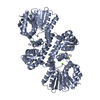 8r4zC  8rprC 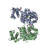 8rvcC 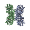 8rvsC 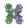 8rwmC 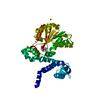 8rwwC 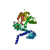 8rxfC C: citing same article ( |
|---|---|
| Similar structure data |
-Links
 PDBj
PDBj
- Assembly
| Deposited unit | 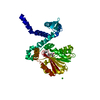
| ||||||||
|---|---|---|---|---|---|---|---|---|---|
| 1 | 
| ||||||||
| Unit cell |
|
-Components
-Protein , 1 types, 1 molecules A
| #1: Protein | Mass: 36911.477 Da / Num. of mol.: 1 Source method: isolated from a genetically manipulated source Source: (gene. exp.) Streptomyces griseoviridis (bacteria) / Plasmid: pET28a (+) / Production host: |
|---|
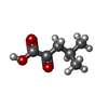
-Non-polymers , 5 types, 352 molecules 


| #2: Chemical | ChemComp-ZN /  Mass: 65.409 Da / Num. of mol.: 1 / Source method: obtained synthetically / Formula: Zn Mass: 65.409 Da / Num. of mol.: 1 / Source method: obtained synthetically / Formula: Zn |
|---|---|
| #3: Chemical | ChemComp-COI /  Mass: 130.142 Da / Num. of mol.: 1 / Source method: obtained synthetically / Formula: C6H10O3 / Feature type: SUBJECT OF INVESTIGATION Mass: 130.142 Da / Num. of mol.: 1 / Source method: obtained synthetically / Formula: C6H10O3 / Feature type: SUBJECT OF INVESTIGATION |
| #4: Chemical | ChemComp-CL /  Mass: 35.453 Da / Num. of mol.: 1 / Source method: obtained synthetically / Formula: Cl Mass: 35.453 Da / Num. of mol.: 1 / Source method: obtained synthetically / Formula: Cl |
| #5: Chemical | ChemComp-SAM /  Mass: 398.437 Da / Num. of mol.: 1 / Source method: obtained synthetically / Formula: C15H22N6O5S Mass: 398.437 Da / Num. of mol.: 1 / Source method: obtained synthetically / Formula: C15H22N6O5S |
| #6: Water | ChemComp-HOH / Mass: 18.015 Da / Num. of mol.: 348 / Source method: isolated from a natural source / Formula: H2O |
-Details
| Has ligand of interest | Y |
|---|---|
| Has protein modification | N |
-Experimental details
-Experiment
| Experiment | Method: X-RAY DIFFRACTION / Number of used crystals: 1 |
|---|
- Sample preparation
| Crystal | Density Matthews: 2.8 Å3/Da / Density % sol: 56.06 % |
|---|---|
| Crystal grow | Temperature: 298 K / Method: vapor diffusion, sitting drop / pH: 8.5 Details: 100 mM Tris/HCl pH 8.5, 250 mM Li2SO4, and 23% PEG 3350 |
-Data collection
| Diffraction | Mean temperature: 100 K / Serial crystal experiment: N |
|---|---|
| Diffraction source | Source: SYNCHROTRON / Site: SLS  / Beamline: X06DA / Wavelength: 1.00004 Å / Beamline: X06DA / Wavelength: 1.00004 Å |
| Detector | Type: DECTRIS PILATUS3 6M / Detector: PIXEL / Date: Mar 13, 2016 |
| Radiation | Protocol: SINGLE WAVELENGTH / Monochromatic (M) / Laue (L): M / Scattering type: x-ray |
| Radiation wavelength | Wavelength: 1.00004 Å / Relative weight: 1 |
| Reflection | Resolution: 1.813→63.0857 Å / Num. obs: 39049 / % possible obs: 100 % / Redundancy: 26.3 % / CC1/2: 0.999 / Rmerge(I) obs: 0.203 / Rpim(I) all: 0.059 / Rsym value: 0.203 / Net I/σ(I): 12.6 |
| Reflection shell | Resolution: 1.813→1.844 Å / Redundancy: 27.5 % / Rmerge(I) obs: 8.708 / Mean I/σ(I) obs: 0.5 / Num. unique obs: 1880 / CC1/2: 0.35 / Rsym value: 8.708 / % possible all: 100 |
- Processing
| Software |
| ||||||||||||||||||||||||||||||||||||||||||||||||||||||||||||||||||
|---|---|---|---|---|---|---|---|---|---|---|---|---|---|---|---|---|---|---|---|---|---|---|---|---|---|---|---|---|---|---|---|---|---|---|---|---|---|---|---|---|---|---|---|---|---|---|---|---|---|---|---|---|---|---|---|---|---|---|---|---|---|---|---|---|---|---|---|
| Refinement | Method to determine structure: MOLECULAR REPLACEMENT / Resolution: 1.813→63.09 Å / Cor.coef. Fo:Fc: 0.945 / Cor.coef. Fo:Fc free: 0.926 / SU R Cruickshank DPI: 0.15 / Cross valid method: THROUGHOUT / SU R Blow DPI: 0.13 / SU Rfree Blow DPI: 0.127 / SU Rfree Cruickshank DPI: 0.12
| ||||||||||||||||||||||||||||||||||||||||||||||||||||||||||||||||||
| Displacement parameters | Biso mean: 53.89 Å2
| ||||||||||||||||||||||||||||||||||||||||||||||||||||||||||||||||||
| Refine analyze | Luzzati coordinate error obs: 0.23 Å | ||||||||||||||||||||||||||||||||||||||||||||||||||||||||||||||||||
| Refinement step | Cycle: LAST / Resolution: 1.813→63.09 Å
| ||||||||||||||||||||||||||||||||||||||||||||||||||||||||||||||||||
| Refine LS restraints |
| ||||||||||||||||||||||||||||||||||||||||||||||||||||||||||||||||||
| LS refinement shell | Resolution: 1.813→1.83 Å
| ||||||||||||||||||||||||||||||||||||||||||||||||||||||||||||||||||
| Refinement TLS params. | Origin x: -0.4658 Å / Origin y: 23.2931 Å / Origin z: -3.5484 Å
| ||||||||||||||||||||||||||||||||||||||||||||||||||||||||||||||||||
| Refinement TLS group |
|
