 Movie
Movie Controller

[English] 日本語
 Yorodumi
- PDB-8rvs: Crystal structure of alpha keto acid C-methyl-transferases MrsA b... -
+ Open data

- Basic information
| Entry | Database: PDB / ID: 8rvs | ||||||
|---|---|---|---|---|---|---|---|
| Title | Crystal structure of alpha keto acid C-methyl-transferases MrsA bound to SAM | ||||||
 Components Components | 2-ketoarginine methyltransferase | ||||||
 Keywords Keywords | TRANSFERASE / S adenosylmethionine-dependent methyltransferases / biocatalysis / C-alkylation / asymmetric methylation / mutagenesis | ||||||
| Function / homology | 5-guanidino-2-oxopentanoate (3R)-methyltransferase / 2-ketoarginine methyltransferase / S-adenosylmethionine-dependent methyltransferase activity / antibiotic biosynthetic process / methylation / S-adenosyl-L-methionine-dependent methyltransferase superfamily / DI(HYDROXYETHYL)ETHER / S-ADENOSYLMETHIONINE / 2-ketoarginine methyltransferase Function and homology information Function and homology information | ||||||
| Biological species |  Pseudomonas syringae (bacteria) Pseudomonas syringae (bacteria) | ||||||
| Method |  X-RAY DIFFRACTION / SYNCHROTRON / MOLECULAR REPLACEMENT / Resolution: 1.632 Å X-RAY DIFFRACTION / SYNCHROTRON / MOLECULAR REPLACEMENT / Resolution: 1.632 Å | ||||||
 Authors Authors | Gerhardt, S. / Andexer, J.N. | ||||||
| Funding support | 1items
| ||||||
 Citation Citation | Journal: Chembiochem / Year: 2024 Title: Structures and Protein Engineering of the alpha-Keto Acid C-Methyltransferases SgvM and MrsA for Rational Substrate Transfer. Authors: Sommer-Kamann, C. / Breiltgens, J. / Zou, Z. / Gerhardt, S. / Saleem-Batcha, R. / Kemper, F. / Einsle, O. / Andexer, J.N. / Muller, M. | ||||||
| History |
|
- Structure visualization
| Structure viewer | Molecule: MolmilJmol/JSmol |
|---|
- Downloads & links
-Download
| PDBx/mmCIF format | 8rvs.cif.gz | 289 KB | Display | PDBx/mmCIF format |
|---|---|---|---|---|
| PDB format | pdb8rvs.ent.gz | 231.1 KB | Display | PDB format |
| PDBx/mmJSON format | 8rvs.json.gz | Tree view | PDBx/mmJSON format | |
| Others |  Other downloads Other downloads |
-Validation report
| Arichive directory | https://data.pdbj.org/pub/pdb/validation_reports/rv/8rvsftp://data.pdbj.org/pub/pdb/validation_reports/rv/8rvs | HTTPS FTP |
|---|
-Related structure data
| Related structure data |  8r4zC  8rprC  8rvcC  8rwmC  8rwwC 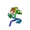 8rxfC 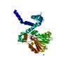 8rxgC C: citing same article ( |
|---|---|
| Similar structure data |
-Links
 PDBj
PDBj
- Assembly
| Deposited unit | 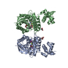
| ||||||||
|---|---|---|---|---|---|---|---|---|---|
| 1 | 
| ||||||||
| Unit cell |
|
-Components
-Protein , 1 types, 2 molecules AB
| #1: Protein | Mass: 38773.242 Da / Num. of mol.: 2 Source method: isolated from a genetically manipulated source Source: (gene. exp.) Pseudomonas syringae (bacteria) / Gene: mrsA / Plasmid: pET-28b (+) / Production host: |
|---|
-Non-polymers , 6 types, 730 molecules 


| #2: Chemical |  Mass: 24.305 Da / Num. of mol.: 2 / Source method: obtained synthetically / Formula: Mg Mass: 24.305 Da / Num. of mol.: 2 / Source method: obtained synthetically / Formula: Mg#3: Chemical |  Mass: 398.437 Da / Num. of mol.: 2 / Source method: obtained synthetically / Formula: C15H22N6O5S / Feature type: SUBJECT OF INVESTIGATION Mass: 398.437 Da / Num. of mol.: 2 / Source method: obtained synthetically / Formula: C15H22N6O5S / Feature type: SUBJECT OF INVESTIGATION#4: Chemical | ChemComp-NA /  Mass: 22.990 Da / Num. of mol.: 4 / Source method: obtained synthetically / Formula: Na Mass: 22.990 Da / Num. of mol.: 4 / Source method: obtained synthetically / Formula: Na#5: Chemical |  Mass: 106.120 Da / Num. of mol.: 2 / Source method: obtained synthetically / Formula: C4H10O3 Mass: 106.120 Da / Num. of mol.: 2 / Source method: obtained synthetically / Formula: C4H10O3#6: Chemical | ChemComp-EDO / |  Mass: 62.068 Da / Num. of mol.: 1 / Source method: obtained synthetically / Formula: C2H6O2 Mass: 62.068 Da / Num. of mol.: 1 / Source method: obtained synthetically / Formula: C2H6O2#7: Water | ChemComp-HOH / | Mass: 18.015 Da / Num. of mol.: 719 / Source method: isolated from a natural source / Formula: H2O |
|---|
-Details
| Has ligand of interest | Y |
|---|---|
| Has protein modification | N |
-Experimental details
-Experiment
| Experiment | Method: X-RAY DIFFRACTION / Number of used crystals: 1 |
|---|
- Sample preparation
| Crystal | Density Matthews: 2.33 Å3/Da / Density % sol: 47.1 % |
|---|---|
| Crystal grow | Temperature: 293 K / Method: vapor diffusion, sitting drop / pH: 7.5 / Details: 100 mM HEPES pH 7.5, 300 mM NaCl, 28% PEG 3350 |
-Data collection
| Diffraction | Mean temperature: 100 K / Serial crystal experiment: N |
|---|---|
| Diffraction source | Source: SYNCHROTRON / Site: SLS  / Beamline: X06SA / Wavelength: 1 Å / Beamline: X06SA / Wavelength: 1 Å |
| Detector | Type: DECTRIS PILATUS 6M-F / Detector: PIXEL / Date: Jun 14, 2015 |
| Radiation | Protocol: SINGLE WAVELENGTH / Monochromatic (M) / Laue (L): M / Scattering type: x-ray |
| Radiation wavelength | Wavelength: 1 Å / Relative weight: 1 |
| Reflection | Resolution: 1.632→66.533 Å / Num. obs: 90343 / % possible obs: 99.7 % / Redundancy: 4.4 % / CC1/2: 0.997 / Rmerge(I) obs: 0.099 / Rpim(I) all: 0.053 / Rrim(I) all: 0.113 / Net I/σ(I): 8.5 |
| Reflection shell | Resolution: 1.632→1.66 Å / Redundancy: 4.5 % / Rmerge(I) obs: 1.574 / Num. unique obs: 4489 / CC1/2: 0.389 / Rpim(I) all: 0.837 / % possible all: 99.9 |
- Processing
| Software |
| |||||||||||||||||||||||||||||||||||||||||||||||||||||||||||||||||||||||||||
|---|---|---|---|---|---|---|---|---|---|---|---|---|---|---|---|---|---|---|---|---|---|---|---|---|---|---|---|---|---|---|---|---|---|---|---|---|---|---|---|---|---|---|---|---|---|---|---|---|---|---|---|---|---|---|---|---|---|---|---|---|---|---|---|---|---|---|---|---|---|---|---|---|---|---|---|---|
| Refinement | Method to determine structure: MOLECULAR REPLACEMENT / Resolution: 1.632→66.53 Å / Cor.coef. Fo:Fc: 0.96 / Cor.coef. Fo:Fc free: 0.952 / SU R Cruickshank DPI: 0.091 / Cross valid method: THROUGHOUT / SU R Blow DPI: 0.098 / SU Rfree Blow DPI: 0.093 / SU Rfree Cruickshank DPI: 0.09
| |||||||||||||||||||||||||||||||||||||||||||||||||||||||||||||||||||||||||||
| Displacement parameters | Biso mean: 28.17 Å2
| |||||||||||||||||||||||||||||||||||||||||||||||||||||||||||||||||||||||||||
| Refine analyze | Luzzati coordinate error obs: 0.21 Å | |||||||||||||||||||||||||||||||||||||||||||||||||||||||||||||||||||||||||||
| Refinement step | Cycle: LAST / Resolution: 1.632→66.53 Å
| |||||||||||||||||||||||||||||||||||||||||||||||||||||||||||||||||||||||||||
| Refine LS restraints |
| |||||||||||||||||||||||||||||||||||||||||||||||||||||||||||||||||||||||||||
| LS refinement shell | Resolution: 1.64→1.66 Å
| |||||||||||||||||||||||||||||||||||||||||||||||||||||||||||||||||||||||||||
| Refinement TLS params. | Refine-ID: X-RAY DIFFRACTION
| |||||||||||||||||||||||||||||||||||||||||||||||||||||||||||||||||||||||||||
| Refinement TLS group |
|
