- PDB-8ru8: A crystal form of a human CDK2-CDK7 chimera -
+
Open data
ID or keywords:
Loading...
-
Basic information
Entry
Database: PDB / ID: 8ru8
Title
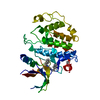
A crystal form of a human CDK2-CDK7 chimera
Components
Cyclin-dependent kinase 2
Keywords
SIGNALING PROTEIN / cyclin dependent kinase 7 / serine/threonine kinase / ATP binding / cell cycle kinase
Function / homology
Function and homology information
cyclin A1-CDK2 complex / cyclin E2-CDK2 complex / cyclin E1-CDK2 complex / cyclin A2-CDK2 complex / positive regulation of DNA-templated DNA replication initiation / G2 Phase / Y chromosome / cyclin-dependent protein kinase activity / regulation of heterochromatin organization / Phosphorylation of proteins involved in G1/S transition by active Cyclin E:Cdk2 complexes ...cyclin A1-CDK2 complex / cyclin E2-CDK2 complex / cyclin E1-CDK2 complex / cyclin A2-CDK2 complex / positive regulation of DNA-templated DNA replication initiation / G2 Phase / Y chromosome / cyclin-dependent protein kinase activity / regulation of heterochromatin organization / Phosphorylation of proteins involved in G1/S transition by active Cyclin E:Cdk2 complexes / positive regulation of heterochromatin formation / p53-Dependent G1 DNA Damage Response / X chromosome / PTK6 Regulates Cell Cycle / regulation of anaphase-promoting complex-dependent catabolic process / Defective binding of RB1 mutants to E2F1,(E2F2, E2F3) / centriole replication / Regulation of APC/C activators between G1/S and early anaphase / telomere maintenance in response to DNA damage / centrosome duplication / G0 and Early G1 / Telomere Extension By Telomerase / Activation of the pre-replicative complex / cyclin-dependent kinase / cyclin-dependent protein serine/threonine kinase activity / TP53 Regulates Transcription of Genes Involved in G1 Cell Cycle Arrest / Activation of ATR in response to replication stress / Regulation of MITF-M-dependent genes involved in cell cycle and proliferation / Cajal body / Cyclin E associated events during G1/S transition / Cyclin A:Cdk2-associated events at S phase entry / cyclin-dependent protein kinase holoenzyme complex / Cyclin A/B1/B2 associated events during G2/M transition / regulation of G2/M transition of mitotic cell cycle / condensed chromosome / mitotic G1 DNA damage checkpoint signaling / negative regulation of protein localization to chromatin / cellular response to nitric oxide / post-translational protein modification / regulation of mitotic cell cycle / cyclin binding / positive regulation of DNA replication / peptidyl-serine phosphorylation / male germ cell nucleus / meiotic cell cycle / potassium ion transport / G1/S transition of mitotic cell cycle / DNA Damage/Telomere Stress Induced Senescence / Meiotic recombination / G2/M transition of mitotic cell cycle / CDK-mediated phosphorylation and removal of Cdc6 / Transcriptional regulation of granulopoiesis / SCF(Skp2)-mediated degradation of p27/p21 / Orc1 removal from chromatin / cellular senescence / Cyclin D associated events in G1 / Regulation of TP53 Degradation / nuclear envelope / Factors involved in megakaryocyte development and platelet production / regulation of gene expression / Processing of DNA double-strand break ends / Senescence-Associated Secretory Phenotype (SASP) / transcription regulator complex / Regulation of TP53 Activity through Phosphorylation / protein phosphorylation / Ras protein signal transduction / DNA replication / chromosome, telomeric region / endosome / chromatin remodeling / protein domain specific binding / protein serine kinase activity / cell division / DNA repair / protein serine/threonine kinase activity / positive regulation of cell population proliferation / centrosome / positive regulation of DNA-templated transcription / DNA-templated transcription / magnesium ion binding / negative regulation of transcription by RNA polymerase II / signal transduction / nucleoplasm / ATP binding / nucleus / cytoplasm / cytosol Similarity search - Function
: / Serine/threonine-protein kinase, active site / Serine/Threonine protein kinases active-site signature. / Protein kinase domain / Serine/Threonine protein kinases, catalytic domain / Protein kinase, ATP binding site / Protein kinases ATP-binding region signature. / Protein kinase domain profile. / Protein kinase domain / Protein kinase-like domain superfamily Similarity search - Domain/homology
Protocol: SINGLE WAVELENGTH / Monochromatic (M) / Laue (L): M / Scattering type: x-ray
Radiation wavelength
Wavelength: 0.9999 Å / Relative weight: 1
Reflection
Resolution: 1.51→51.17 Å / Num. obs: 45158 / % possible obs: 99.7 % / Redundancy: 6.18 % / Rmerge(I) obs: 0.085 / Net I/σ(I): 10.1
Reflection shell
Resolution: 1.51→1.536 Å / Mean I/σ(I) obs: 1.1 / Num. unique obs: 2202 / Rrim(I) all: 1.492 / % possible all: 99.1
-
Processing
Software
Name
Version
Classification
BUSTER
2.11.8 (16-JUL-2021)
refinement
X-Area
datareduction
Aimless
datascaling
BUSTER
phasing
Refinement
Method to determine structure: FOURIER SYNTHESIS / Resolution: 1.51→51.17 Å / Cor.coef. Fo:Fc: 0.944 / Cor.coef. Fo:Fc free: 0.923 / SU R Cruickshank DPI: 0.117 / Cross valid method: THROUGHOUT / σ(F): 0 / SU R Blow DPI: 0.102 / SU Rfree Blow DPI: 0.102 / SU Rfree Cruickshank DPI: 0.098 Details: HYDROGENS WERE FULLY REFINED WITH FULL OCCUPANCY AT NUCLEAR POSITION. REFINEMENT NOTES. TYPES WITHOUT CCP4 ATOM TYPE IN LIBRARY=. NUMBER OF ATOMS WITH PROPER CCP4 ATOM TYPE=4921. NUMBER WITH ...Details: HYDROGENS WERE FULLY REFINED WITH FULL OCCUPANCY AT NUCLEAR POSITION. REFINEMENT NOTES. TYPES WITHOUT CCP4 ATOM TYPE IN LIBRARY=. NUMBER OF ATOMS WITH PROPER CCP4 ATOM TYPE=4921. NUMBER WITH APPROX DEFAULT CCP4 ATOM TYPE=0. NUMBER TREATED BY BAD NON-BONDED CONTACTS=3.
Rfactor
Num. reflection
% reflection
Selection details
Rfree
0.2408
1995
5.07 %
RANDOM
Rwork
0.2033
-
-
-
obs
0.2051
39316
86.8 %
-
Displacement parameters
Biso mean: 28.809 Å2
Baniso -1
Baniso -2
Baniso -3
1-
4.167 Å2
0 Å2
0 Å2
2-
-
1.5436 Å2
0 Å2
3-
-
-
-5.7107 Å2
Refine analyze
Luzzati coordinate error obs: 0.22 Å
Refinement step
Cycle: LAST / Resolution: 1.51→51.17 Å
Protein
Nucleic acid
Ligand
Solvent
Total
Num. atoms
2227
0
29
290
2546
Refine LS restraints
Refine-ID
Type
Dev ideal
Number
Restraint function
Weight
X-RAY DIFFRACTION
t_bond_d
0.014
4695
HARMONIC
2
X-RAY DIFFRACTION
t_angle_deg
1.18
8534
HARMONIC
2
X-RAY DIFFRACTION
t_dihedral_angle_d
1028
SINUSOIDAL
2
X-RAY DIFFRACTION
t_incorr_chiral_ct
X-RAY DIFFRACTION
t_pseud_angle
X-RAY DIFFRACTION
t_trig_c_planes
X-RAY DIFFRACTION
t_gen_planes
735
HARMONIC
16
X-RAY DIFFRACTION
t_it
4695
HARMONIC
10
X-RAY DIFFRACTION
t_nbd
4
SEMIHARMONIC
5
X-RAY DIFFRACTION
t_omega_torsion
6.11
X-RAY DIFFRACTION
t_other_torsion
17.59
X-RAY DIFFRACTION
t_improper_torsion
X-RAY DIFFRACTION
t_chiral_improper_torsion
296
SEMIHARMONIC
5
X-RAY DIFFRACTION
t_sum_occupancies
X-RAY DIFFRACTION
t_utility_distance
X-RAY DIFFRACTION
t_utility_angle
X-RAY DIFFRACTION
t_utility_torsion
X-RAY DIFFRACTION
t_ideal_dist_contact
4008
SEMIHARMONIC
4
LS refinement shell
Resolution: 1.51→1.53 Å / Total num. of bins used: 51
Rfactor
Num. reflection
% reflection
Rfree
0.32
-
4.45 %
Rwork
0.2698
752
-
all
0.2719
787
-
obs
-
-
41.24 %
Refinement TLS params.
Method: refined / Refine-ID: X-RAY DIFFRACTION
ID
L11 (°2)
L12 (°2)
L13 (°2)
L22 (°2)
L23 (°2)
L33 (°2)
S11 (Å °)
S12 (Å °)
S13 (Å °)
S21 (Å °)
S22 (Å °)
S23 (Å °)
S31 (Å °)
S32 (Å °)
S33 (Å °)
T11 (Å2)
T12 (Å2)
T13 (Å2)
T22 (Å2)
T23 (Å2)
T33 (Å2)
Origin x (Å)
Origin y (Å)
Origin z (Å)
1
0.6515
-0.5375
-0.5433
0.8543
0.8106
3.9339
-0.0287
-0.0341
-0.1251
-0.0004
-0.1111
0.2217
-0.0734
-0.5343
0.1398
-0.0004
-0.0127
0.0059
0.0962
0.03
-0.0366
18.9316
2.0137
65.804
2
0.4562
-0.5574
0.7979
0
-0.2505
2.284
0.0829
-0.1316
0.0435
0.1265
-0.0272
0.0385
-0.1027
-0.099
-0.0557
0.0618
-0.0004
0.0132
0.0422
0.0229
-0.079
30.4235
5.3247
57.3044
3
0.2324
-0.3645
0.2531
0.1128
0.1674
1.2886
0.0655
0.0165
0.013
-0.0261
-0.0429
0.0106
-0.0733
0.0736
-0.0225
0.0477
0.0011
0.0009
0.012
0.0157
-0.076
33.8568
6.5969
42.723
Refinement TLS group
ID
Refine-ID
Refine TLS-ID
Selection details
1
X-RAY DIFFRACTION
1
{ A|1 - A|83 }
2
X-RAY DIFFRACTION
2
{ A|130 - A|164 }
3
X-RAY DIFFRACTION
3
{ A|84 - A|129 A|165 - A|298 }
+
About Yorodumi
-
News
-
Feb 9, 2022. New format data for meta-information of EMDB entries
New format data for meta-information of EMDB entries
Version 3 of the EMDB header file is now the official format.
The previous official version 1.9 will be removed from the archive.
In the structure databanks used in Yorodumi, some data are registered as the other names, "COVID-19 virus" and "2019-nCoV". Here are the details of the virus and the list of structure data.
Jan 31, 2019. EMDB accession codes are about to change! (news from PDBe EMDB page)
EMDB accession codes are about to change! (news from PDBe EMDB page)
The allocation of 4 digits for EMDB accession codes will soon come to an end. Whilst these codes will remain in use, new EMDB accession codes will include an additional digit and will expand incrementally as the available range of codes is exhausted. The current 4-digit format prefixed with “EMD-” (i.e. EMD-XXXX) will advance to a 5-digit format (i.e. EMD-XXXXX), and so on. It is currently estimated that the 4-digit codes will be depleted around Spring 2019, at which point the 5-digit format will come into force.
The EM Navigator/Yorodumi systems omit the EMD- prefix.
Related info.:Q: What is EMD? / ID/Accession-code notation in Yorodumi/EM Navigator
Yorodumi is a browser for structure data from EMDB, PDB, SASBDB, etc.
This page is also the successor to EM Navigator detail page, and also detail information page/front-end page for Omokage search.
The word "yorodu" (or yorozu) is an old Japanese word meaning "ten thousand". "mi" (miru) is to see.
Related info.:EMDB / PDB / SASBDB / Comparison of 3 databanks / Yorodumi Search / Aug 31, 2016. New EM Navigator & Yorodumi / Yorodumi Papers / Jmol/JSmol / Function and homology information / Changes in new EM Navigator and Yorodumi
 Movie
Movie Controller


 Open data
 Basic information
 Components
 Keywords
 Function and homology information
 Homo sapiens (human)
 X-RAY DIFFRACTION /
  Authors
 Citation
 Structure visualization
 Downloads & links
 Other downloads


 PDBj
PDBj


 Assembly
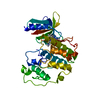


 Mass: 394.513 Da / Num. of mol.: 1 / Source method: obtained synthetically / Formula: C22H30N6O / Feature type: SUBJECT OF INVESTIGATION
 Mass: 18.015 Da / Num. of mol.: 290 / Source method: isolated from a natural source / Formula: H2O
 Sample preparation
 / Beamline: I24 / Wavelength: 0.9999 Å
 Processing