[English] 日本語
Yorodumi
- PDB-8rqz: Crystal structure of Molybdenum bispyranopterin guanine dinucleot... -

+
Open data


ID or keywords:

Loading...

-
Basic information

Entry
Database: PDB / ID: 8rqz
TitleCrystal structure of Molybdenum bispyranopterin guanine dinucleotide formate dehydrogenases ForCE1 from Bacillus subtilis
Components
  • Probable oxidoreductase YjgC
  • Uncharacterized protein YjgD
KeywordsOXIDOREDUCTASE / Bacterial metabolism Bioenergetics Metalloenzyme Quinone Iron-Sulfur Cluster Helical Membrane Plug-in
Function / homology
Function and homology information


formate metabolic process / formate dehydrogenase (NAD+) activity / Oxidoreductases / molybdopterin cofactor binding / respiratory electron transport chain / 2 iron, 2 sulfur cluster binding / 4 iron, 4 sulfur cluster binding / metal ion binding / membrane
Similarity search - Function
Protein of unknown function DUF1641 / Protein of unknown function (DUF1641) / Formate dehydrogenase, alpha subunit / Formate dehydrogenase H, N-terminal / Molybdopterin oxidoreductase Fe4S4 domain / Molybdopterin oxidoreductase Fe4S4 domain / Molybdopterin dinucleotide-binding domain / Molydopterin dinucleotide binding domain / : / NADH-ubiquinone oxidoreductase-G iron-sulfur binding region ...Protein of unknown function DUF1641 / Protein of unknown function (DUF1641) / Formate dehydrogenase, alpha subunit / Formate dehydrogenase H, N-terminal / Molybdopterin oxidoreductase Fe4S4 domain / Molybdopterin oxidoreductase Fe4S4 domain / Molybdopterin dinucleotide-binding domain / Molydopterin dinucleotide binding domain / : / NADH-ubiquinone oxidoreductase-G iron-sulfur binding region / NADH-ubiquinone oxidoreductase-G iron-sulfur binding region / 2Fe-2S iron-sulfur cluster binding domain / NADH:ubiquinone oxidoreductase, subunit G, iron-sulphur binding / His(Cys)3-ligated-type [4Fe-4S] domain profile. / Aspartate decarboxylase-like domain superfamily / Molybdopterin oxidoreductase, 4Fe-4S domain / Prokaryotic molybdopterin oxidoreductases 4Fe-4S domain profile. / Molybdopterin oxidoreductase / Molybdopterin oxidoreductase / 4Fe-4S dicluster domain / 2Fe-2S ferredoxin-type iron-sulfur binding domain profile. / 2Fe-2S ferredoxin-type iron-sulfur binding domain / 2Fe-2S ferredoxin-like superfamily / 4Fe-4S ferredoxin, iron-sulphur binding, conserved site / 4Fe-4S ferredoxin-type iron-sulfur binding region signature. / 4Fe-4S ferredoxin-type iron-sulfur binding domain profile. / 4Fe-4S ferredoxin-type, iron-sulphur binding domain
Similarity search - Domain/homology
MOLYBDENUM(IV) ION / FE2/S2 (INORGANIC) CLUSTER / HYDROSULFURIC ACID / 1,2-DIPALMITOYL-PHOSPHATIDYL-GLYCEROLE / Chem-MGD / MALONATE ION / MENAQUINONE-7 / MYRISTIC ACID / NITRATE ION / IRON/SULFUR CLUSTER ...MOLYBDENUM(IV) ION / FE2/S2 (INORGANIC) CLUSTER / HYDROSULFURIC ACID / 1,2-DIPALMITOYL-PHOSPHATIDYL-GLYCEROLE / Chem-MGD / MALONATE ION / MENAQUINONE-7 / MYRISTIC ACID / NITRATE ION / IRON/SULFUR CLUSTER / Uncharacterized protein YjgD / Probable oxidoreductase YjgC
Similarity search - Component
Biological speciesBacillus subtilis subsp. subtilis str. 168 (bacteria)
MethodX-RAY DIFFRACTION / SYNCHROTRON / MOLECULAR REPLACEMENT / Resolution: 2.694 Å
AuthorsCherrier, M.V. / Arnoux, P. / Martin, L. / Nicolet, Y. / Schoehn, G. / Legrand, P. / Broc, M. / Seduk, F. / Brasseur, G. / Arias-Cartin, R. ...Cherrier, M.V. / Arnoux, P. / Martin, L. / Nicolet, Y. / Schoehn, G. / Legrand, P. / Broc, M. / Seduk, F. / Brasseur, G. / Arias-Cartin, R. / Magalon, A. / Walburger, A. / Uzel, A. / Guigliarelli, B. / Grimaldi, S. / Pierrel, F. / Mate, M.
Funding support France, 6items
OrganizationGrant numberCountry
Agence Nationale de la Recherche (ANR)ANR-15-IDEX-02 France
French Infrastructure for Integrated Structural Biology (FRISBI)ANR-10-INBS-05-02 France
Grenoble Alliance for Integrated Structural Cell Biology (GRAL)ANR-17-EURE-0003 France
Agence Nationale de la Recherche (ANR)ANR-22-CE44-0035-01 France
Centre National de la Recherche Scientifique (CNRS)MICROBIOCO2 France
Agence Nationale de la Recherche (ANR)ANR 16-CE29-0010-01 France
CitationJournal: Nat Struct Mol Biol / Year: 2025
Title: A scaffold for quinone channeling between membrane and soluble bacterial oxidoreductases.
Authors: M Broc / M V Cherrier / A Uzel / R Arias-Cartin / P Arnoux / G Brasseur / F Seduk / B Guigliarelli / P Legrand / F Pierrel / G Schoehn / M J Maté / L Martin / S Grimaldi / Y Nicolet / A Magalon / A Walburger /
Abstract: Redox processes are at the heart of energetic metabolism that drives life on earth. By extension, complex and efficient electron transfer wires are necessary to connect the various metabolic pathways ...Redox processes are at the heart of energetic metabolism that drives life on earth. By extension, complex and efficient electron transfer wires are necessary to connect the various metabolic pathways that are often located in distinct cellular compartments. Here, we uncovered a structural module that enables channeling of quinones from the membrane to various water-soluble redox catalytic units in prokaryotes. Using X-ray crystallography and cryo-electron microscopy, we determined the structure of the unusual bacterial formate dehydrogenase ForCE that contains four ForC catalytic subunits docked around a membrane-associated tetrameric ForE central scaffold. In the latter, a conserved domain that we propose to name helical membrane plugin (HMP) was identified as essential to link formate oxidation, in Bacillus subtilis, to the aerobic respiratory chain. Our bioinformatic analysis indicates that this HMP is associated with different quinone-reducing oxidoreductases, highlighting its broad importance as a functional unit to wire electrons between a given catalytic redox center and the quinone pool.
History
DepositionJan 22, 2024Deposition site: PDBE / Processing site: PDBE
Revision 1.0Aug 6, 2025Provider: repository / Type: Initial release
Revision 1.1Aug 27, 2025Group: Database references / Category: citation
Item: _citation.country / _citation.journal_abbrev ..._citation.country / _citation.journal_abbrev / _citation.journal_id_CSD / _citation.journal_id_ISSN / _citation.pdbx_database_id_DOI / _citation.title / _citation.year
Revision 1.2Sep 3, 2025Group: Database references / Category: citation / citation_author
Item: _citation.pdbx_database_id_PubMed / _citation.title ..._citation.pdbx_database_id_PubMed / _citation.title / _citation_author.identifier_ORCID / _citation_author.name
Revision 1.3Nov 26, 2025Group: Database references / Category: citation
Item: _citation.journal_volume / _citation.page_first / _citation.page_last

-
Structure visualization

Structure viewerMolecule:
MolmilJmol/JSmol

Downloads & links

-
Assembly

Deposited unit
A: Uncharacterized protein YjgD
B: Uncharacterized protein YjgD
C: Probable oxidoreductase YjgC
D: Probable oxidoreductase YjgC
hetero molecules


Theoretical massNumber of molelcules
Total (without water)274,66553
Polymers262,4664
Non-polymers12,19949
Water12,376687
1
A: Uncharacterized protein YjgD
B: Uncharacterized protein YjgD
C: Probable oxidoreductase YjgC
D: Probable oxidoreductase YjgC
hetero molecules

A: Uncharacterized protein YjgD
B: Uncharacterized protein YjgD
C: Probable oxidoreductase YjgC
D: Probable oxidoreductase YjgC
hetero molecules


Theoretical massNumber of molelcules
Total (without water)549,331106
Polymers524,9338
Non-polymers24,39898
Water1448
TypeNameSymmetry operationNumber
identity operation1_555x,y,z1
point symmetry operation1
Buried area68670 Å2
ΔGint-875 kcal/mol
Surface area153600 Å2
Unit cell
Length a, b, c (Å)210.833, 210.500, 494.213
Angle α, β, γ (deg.)90.00, 90.00, 90.00
Int Tables number22
Space group name H-MF222

-
Components

-
Protein , 2 types, 4 molecules ABCD

#1: Protein Uncharacterized protein YjgD


Mass: 21308.650 Da / Num. of mol.: 2 / Source method: isolated from a natural source
Source: (natural) Bacillus subtilis subsp. subtilis str. 168 (bacteria)
References: UniProt: O34681
#2: Protein Probable oxidoreductase YjgC


Mass: 109924.477 Da / Num. of mol.: 2 / Source method: isolated from a natural source
Source: (natural) Bacillus subtilis subsp. subtilis str. 168 (bacteria)
References: UniProt: O34720, Oxidoreductases

-
Non-polymers , 13 types, 736 molecules

#3: Chemical
ChemComp-MYR / MYRISTIC ACID


Mass: 228.371 Da / Num. of mol.: 5 / Source method: obtained synthetically / Formula: C14H28O2
#4: Chemical
ChemComp-GOL / GLYCEROL / GLYCERIN / PROPANE-1,2,3-TRIOL


Mass: 92.094 Da / Num. of mol.: 8 / Source method: obtained synthetically / Formula: C3H8O3
#5: Chemical ChemComp-LHG / 1,2-DIPALMITOYL-PHOSPHATIDYL-GLYCEROLE


Mass: 722.970 Da / Num. of mol.: 2 / Source method: obtained synthetically / Formula: C38H75O10P / Comment: phospholipid*YM
#6: Chemical ChemComp-FES / FE2/S2 (INORGANIC) CLUSTER


Mass: 175.820 Da / Num. of mol.: 2 / Source method: obtained synthetically / Formula: Fe2S2 / Feature type: SUBJECT OF INVESTIGATION
#7: Chemical
ChemComp-SF4 / IRON/SULFUR CLUSTER


Mass: 351.640 Da / Num. of mol.: 8 / Source method: obtained synthetically / Formula: Fe4S4 / Feature type: SUBJECT OF INVESTIGATION
#8: Chemical
ChemComp-MGD / 2-AMINO-5,6-DIMERCAPTO-7-METHYL-3,7,8A,9-TETRAHYDRO-8-OXA-1,3,9,10-TETRAAZA-ANTHRACEN-4-ONE GUANOSINE DINUCLEOTIDE / MOLYBDOPTERIN GUANOSINE DINUCLEOTIDE


Mass: 740.557 Da / Num. of mol.: 4 / Source method: obtained synthetically / Formula: C20H26N10O13P2S2
#9: Chemical ChemComp-4MO / MOLYBDENUM(IV) ION


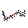
Mass: 95.940 Da / Num. of mol.: 2 / Source method: obtained synthetically / Formula: Mo / Feature type: SUBJECT OF INVESTIGATION
#10: Chemical ChemComp-MQ7 / MENAQUINONE-7


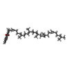
Mass: 648.999 Da / Num. of mol.: 2 / Source method: obtained synthetically / Formula: C46H64O2 / Feature type: SUBJECT OF INVESTIGATION
#11: Chemical ChemComp-H2S / HYDROSULFURIC ACID / HYDROGEN SULFIDE


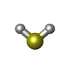
Mass: 34.081 Da / Num. of mol.: 2 / Source method: obtained synthetically / Formula: H2S / Feature type: SUBJECT OF INVESTIGATION
#12: Chemical
ChemComp-MLI / MALONATE ION


Mass: 102.046 Da / Num. of mol.: 9 / Source method: obtained synthetically / Formula: C3H2O4
#13: Chemical
ChemComp-NO3 / NITRATE ION


Mass: 62.005 Da / Num. of mol.: 4 / Source method: obtained synthetically / Formula: NO3
#14: Chemical ChemComp-NA / SODIUM ION


Mass: 22.990 Da / Num. of mol.: 1 / Source method: obtained synthetically / Formula: Na
#15: Water ChemComp-HOH / water


Mass: 18.015 Da / Num. of mol.: 687 / Source method: isolated from a natural source / Formula: H2O

-
Details

Has ligand of interestY
Has protein modificationN

-
Experimental details

-
Experiment

ExperimentMethod: X-RAY DIFFRACTION / Number of used crystals: 1

-
Sample preparation

Crystal growTemperature: 298 K / Method: vapor diffusion, sitting drop / pH: 7 / Details: 1.5 M sodium manolate pH 7

-
Data collection

DiffractionMean temperature: 100 K / Serial crystal experiment: N
Diffraction sourceSource: SYNCHROTRON / Site: ESRF / Beamline: ID23-2 / Wavelength: 0.87313 Å
DetectorType: DECTRIS PILATUS3 X 2M / Detector: PIXEL / Date: Nov 16, 2022
RadiationProtocol: SINGLE WAVELENGTH / Monochromatic (M) / Laue (L): M / Scattering type: x-ray
Radiation wavelengthWavelength: 0.87313 Å / Relative weight: 1
ReflectionResolution: 2.65→49.5 Å / Num. obs: 96030 / % possible obs: 93.6 % / Redundancy: 7.8 % / CC1/2: 0.984 / Rrim(I) all: 0.34 / Net I/σ(I): 5.6
Reflection shellResolution: 2.65→2.88 Å / Redundancy: 7.1 % / Mean I/σ(I) obs: 1.5 / Num. unique obs: 34385 / CC1/2: 0.533 / Rrim(I) all: 0.1363 / % possible all: 65.8

-
Processing

Software
NameVersionClassification
BUSTER2.10.4 (26-JUL-2023)refinement
XDSJan 10, 2022data reduction
STARANISO2.3.89data scaling
PHASER2.8.3phasing
RefinementMethod to determine structure: MOLECULAR REPLACEMENT / Resolution: 2.694→49.41 Å / Cor.coef. Fo:Fc: 0.888 / Cor.coef. Fo:Fc free: 0.856 / SU R Cruickshank DPI: 0.657 / Cross valid method: THROUGHOUT / σ(F): 0 / SU R Blow DPI: 0.654 / SU Rfree Blow DPI: 0.325 / SU Rfree Cruickshank DPI: 0.329
RfactorNum. reflection% reflectionSelection details
Rfree0.2306 4688 5.07 %RANDOM
Rwork0.1956 ---
obs0.1974 92424 61.5 %-
Displacement parametersBiso mean: 59.56 Å2
Baniso -1Baniso -2Baniso -3
1--10.5994 Å20 Å20 Å2
2---12.8272 Å20 Å2
3---23.4265 Å2
Refine analyzeLuzzati coordinate error obs: 0.35 Å
Refinement stepCycle: LAST / Resolution: 2.694→49.41 Å
ProteinNucleic acidLigandSolventTotal
Num. atoms17585 0 604 687 18876
Refine LS restraints
Refine-IDTypeDev idealNumberRestraint functionWeight
X-RAY DIFFRACTIONt_bond_d0.00718621HARMONIC2
X-RAY DIFFRACTIONt_angle_deg0.9125222HARMONIC2
X-RAY DIFFRACTIONt_dihedral_angle_d6637SINUSOIDAL2
X-RAY DIFFRACTIONt_incorr_chiral_ct
X-RAY DIFFRACTIONt_pseud_angle
X-RAY DIFFRACTIONt_trig_c_planes
X-RAY DIFFRACTIONt_gen_planes3159HARMONIC5
X-RAY DIFFRACTIONt_it18621HARMONIC10
X-RAY DIFFRACTIONt_nbd
X-RAY DIFFRACTIONt_omega_torsion2.69
X-RAY DIFFRACTIONt_other_torsion19.09
X-RAY DIFFRACTIONt_improper_torsion
X-RAY DIFFRACTIONt_chiral_improper_torsion2389SEMIHARMONIC5
X-RAY DIFFRACTIONt_sum_occupancies8HARMONIC1
X-RAY DIFFRACTIONt_utility_distance52HARMONIC1
X-RAY DIFFRACTIONt_utility_angle
X-RAY DIFFRACTIONt_utility_torsion
X-RAY DIFFRACTIONt_ideal_dist_contact16891SEMIHARMONIC4
LS refinement shellResolution: 2.694→2.83 Å
RfactorNum. reflection% reflection
Rfree0.4444 -3.89 %
Rwork0.2906 1777 -
obs--8.83 %
Refinement TLS params.

Method: refined / Refine-ID: X-RAY DIFFRACTION

IDL112)L122)L132)L222)L232)L332)S11 (Å °)S12 (Å °)S13 (Å °)S21 (Å °)S22 (Å °)S23 (Å °)S31 (Å °)S32 (Å °)S33 (Å °)T112)T122)T132)T222)T232)T332)Origin x (Å)Origin y (Å)Origin z (Å)
10.55830.1326-0.430.4138-0.05090.2741-0.01620.06610.0022-0.0646-0.04440.0959-0.00250.10430.0606-0.00920.02230.0552-0.0911-0.01310.057149.106953.0195180.671
20.2607-0.08550.05230.6091-0.4820.3848-0.03260.0519-0.1806-0.07490.00560.0912-0.01320.00750.0269-0.0521-0.0141-0.0085-0.01050.02720.0352.382549.0101180.104
31.5118-0.2573-0.10740.6543-0.32880.3048-0.0044-0.1056-0.8393-0.1487-0.03760.0931-0.250.05080.0419-0.2884-0.00610.0126-0.28440.09690.46761.13962.6143205.048
40.64390.2338-0.36261.50830.12450.2332-0.02660.1740.08350.1251-0.02410.8760.06560.30250.0508-0.25530.00290.1082-0.3296-0.00830.4822.519244.1884205.092
Refinement TLS group
IDRefine-IDRefine TLS-IDSelection details
1X-RAY DIFFRACTION1{ A|* }
2X-RAY DIFFRACTION2{ B|* }
3X-RAY DIFFRACTION3{ C|* }
4X-RAY DIFFRACTION4{ D|* }

+
About Yorodumi

-
News

-
Feb 9, 2022. New format data for meta-information of EMDB entries

New format data for meta-information of EMDB entries

  • Version 3 of the EMDB header file is now the official format.
  • The previous official version 1.9 will be removed from the archive.

Related info.:EMDB header

External links:wwPDB to switch to version 3 of the EMDB data model

-
Aug 12, 2020. Covid-19 info

Covid-19 info

URL: https://pdbj.org/emnavi/covid19.php

New page: Covid-19 featured information page in EM Navigator.

Related info.:Covid-19 info / Mar 5, 2020. Novel coronavirus structure data

+
Mar 5, 2020. Novel coronavirus structure data

Novel coronavirus structure data

Related info.:Yorodumi Speices / Aug 12, 2020. Covid-19 info

External links:COVID-19 featured content - PDBj / Molecule of the Month (242):Coronavirus Proteases

+
Jan 31, 2019. EMDB accession codes are about to change! (news from PDBe EMDB page)

EMDB accession codes are about to change! (news from PDBe EMDB page)

  • The allocation of 4 digits for EMDB accession codes will soon come to an end. Whilst these codes will remain in use, new EMDB accession codes will include an additional digit and will expand incrementally as the available range of codes is exhausted. The current 4-digit format prefixed with “EMD-” (i.e. EMD-XXXX) will advance to a 5-digit format (i.e. EMD-XXXXX), and so on. It is currently estimated that the 4-digit codes will be depleted around Spring 2019, at which point the 5-digit format will come into force.
  • The EM Navigator/Yorodumi systems omit the EMD- prefix.

Related info.:Q: What is EMD? / ID/Accession-code notation in Yorodumi/EM Navigator

External links:EMDB Accession Codes are Changing Soon! / Contact to PDBj

+
Jul 12, 2017. Major update of PDB

Major update of PDB

  • wwPDB released updated PDB data conforming to the new PDBx/mmCIF dictionary.
  • This is a major update changing the version number from 4 to 5, and with Remediation, in which all the entries are updated.
  • In this update, many items about electron microscopy experimental information are reorganized (e.g. em_software).
  • Now, EM Navigator and Yorodumi are based on the updated data.

External links:wwPDB Remediation / Enriched Model Files Conforming to OneDep Data Standards Now Available in the PDB FTP Archive

-
Yorodumi

Thousand views of thousand structures

  • Yorodumi is a browser for structure data from EMDB, PDB, SASBDB, etc.
  • This page is also the successor to EM Navigator detail page, and also detail information page/front-end page for Omokage search.
  • The word "yorodu" (or yorozu) is an old Japanese word meaning "ten thousand". "mi" (miru) is to see.

Related info.:EMDB / PDB / SASBDB / Comparison of 3 databanks / Yorodumi Search / Aug 31, 2016. New EM Navigator & Yorodumi / Yorodumi Papers / Jmol/JSmol / Function and homology information / Changes in new EM Navigator and Yorodumi

Read more