Movie
Movie Controller

[English] 日本語
 Yorodumi
- PDB-8rm1: Cryo-EM structure of a Foamy Virus fusion glycoprotein in the pos... -
+ Open data

- Basic information
| Entry | Database: PDB / ID: 8rm1 | ||||||||||||||||||||||||||||||||||||||||||||||||
|---|---|---|---|---|---|---|---|---|---|---|---|---|---|---|---|---|---|---|---|---|---|---|---|---|---|---|---|---|---|---|---|---|---|---|---|---|---|---|---|---|---|---|---|---|---|---|---|---|---|
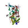
| Title | Cryo-EM structure of a Foamy Virus fusion glycoprotein in the postfusion conformation | ||||||||||||||||||||||||||||||||||||||||||||||||
 Components Components | (Envelope glycoprotein gp130) x 2 | ||||||||||||||||||||||||||||||||||||||||||||||||
 Keywords Keywords | VIRUS / Envelope / Fusion / Spumavirus | ||||||||||||||||||||||||||||||||||||||||||||||||
| Function / homology | Foamy virus envelope protein / Foamy virus envelope protein / host cell endoplasmic reticulum membrane / viral envelope / virion membrane / membrane / Envelope glycoprotein gp130 Function and homology information Function and homology information | ||||||||||||||||||||||||||||||||||||||||||||||||
| Biological species |  Simian foamy virus Simian foamy virus | ||||||||||||||||||||||||||||||||||||||||||||||||
| Method | ELECTRON MICROSCOPY / single particle reconstruction / cryo EM / Resolution: 3.1 Å | ||||||||||||||||||||||||||||||||||||||||||||||||
 Authors Authors | Fernandez, I. / Backovic, M. | ||||||||||||||||||||||||||||||||||||||||||||||||
| Funding support |  France, 1items France, 1items
| ||||||||||||||||||||||||||||||||||||||||||||||||
 Citation Citation | Journal: Sci Adv / Year: 2024 Title: Structures of the Foamy virus fusion protein reveal an unexpected link with the F protein of paramyxo- and pneumoviruses. Authors: Ignacio Fernández / François Bontems / Delphine Brun / Youna Coquin / Casper A Goverde / Bruno E Correia / Antoine Gessain / Florence Buseyne / Felix A Rey / Marija Backovic /  Abstract: Foamy viruses (FVs) constitute a subfamily of retroviruses. Their envelope (Env) glycoprotein drives the merger of viral and cellular membranes during entry into cells. The only available structures ...Foamy viruses (FVs) constitute a subfamily of retroviruses. Their envelope (Env) glycoprotein drives the merger of viral and cellular membranes during entry into cells. The only available structures of retroviral Envs are those from human and simian immunodeficiency viruses from the subfamily of orthoretroviruses, which are only distantly related to the FVs. We report the cryo-electron microscopy structures of the FV Env ectodomain in the pre- and post-fusion states, which unexpectedly demonstrate structural similarity with the fusion protein (F) of paramyxo- and pneumoviruses, implying an evolutionary link between the viral fusogens. We describe the structural features that are unique to the FV Env and propose a mechanistic model for its conformational change, highlighting how the interplay of its structural elements could drive membrane fusion and viral entry. The structural knowledge on the FV Env now provides a framework for functional investigations, which can benefit the design of FV Env variants with improved features for use as gene therapy vectors. | ||||||||||||||||||||||||||||||||||||||||||||||||
| History |
|
- Structure visualization
| Structure viewer | Molecule: MolmilJmol/JSmol |
|---|
- Downloads & links
-Download
| PDBx/mmCIF format | 8rm1.cif.gz | 401.1 KB | Display | PDBx/mmCIF format |
|---|---|---|---|---|
| PDB format | pdb8rm1.ent.gz | 326.5 KB | Display | PDB format |
| PDBx/mmJSON format | 8rm1.json.gz | Tree view | PDBx/mmJSON format | |
| Others |  Other downloads Other downloads |
-Validation report
| Summary document | 8rm1_validation.pdf.gz | 1.7 MB | Display | wwPDB validaton report |
|---|---|---|---|---|
| Full document | 8rm1_full_validation.pdf.gz | 1.7 MB | Display | |
| Data in XML | 8rm1_validation.xml.gz | 66.5 KB | Display | |
| Data in CIF | 8rm1_validation.cif.gz | 100.6 KB | Display | |
| Arichive directory | https://data.pdbj.org/pub/pdb/validation_reports/rm/8rm1ftp://data.pdbj.org/pub/pdb/validation_reports/rm/8rm1 | HTTPS FTP |
-Related structure data
| Related structure data |  19348MC  8rm0C M: map data used to model this data C: citing same article ( |
|---|---|
| Similar structure data |


-Links
 PDBj
PDBj- Assembly
| Deposited unit | 
|
|---|---|
| 1 |
|
-Components
| #1: Protein | Mass: 52050.996 Da / Num. of mol.: 3 Source method: isolated from a genetically manipulated source Source: (gene. exp.) Simian foamy virus / Gene: env / Production host:  #2: Protein | Mass: 42067.801 Da / Num. of mol.: 3 Source method: isolated from a genetically manipulated source Source: (gene. exp.) Simian foamy virus / Gene: env / Production host: #3: Polysaccharide | Source method: isolated from a genetically manipulated source #4: Polysaccharide | Source method: isolated from a genetically manipulated source #5: Sugar |   Type: D-saccharide, beta linking / Mass: 221.208 Da / Num. of mol.: 3 / Source method: obtained synthetically / Formula: C8H15NO6 Type: D-saccharide, beta linking / Mass: 221.208 Da / Num. of mol.: 3 / Source method: obtained synthetically / Formula: C8H15NO6Has ligand of interest | N | Has protein modification | Y | |
|---|
-Experimental details
-Experiment
| Experiment | Method: ELECTRON MICROSCOPY |
|---|---|
| EM experiment | Aggregation state: PARTICLE / 3D reconstruction method: single particle reconstruction |
- Sample preparation
| Component | Name: Gorilla Foamy Virus Envelope glycoprotein / Type: COMPLEX / Entity ID: #1 / Source: RECOMBINANT |
|---|---|
| Molecular weight | Experimental value: NO |
| Source (natural) | Organism: Spumavirus |
| Source (recombinant) | Organism: |
| Buffer solution | pH: 8 |
| Specimen | Embedding applied: NO / Shadowing applied: NO / Staining applied: NO / Vitrification applied: YES |
| Specimen support | Grid type: Quantifoil R1.2/1.3 |
| Vitrification | Instrument: FEI VITROBOT MARK IV / Cryogen name: ETHANE / Humidity: 100 % / Chamber temperature: 281 K |
- Electron microscopy imaging
| Experimental equipment |  Model: Titan Krios / Image courtesy: FEI Company |
|---|---|
| Microscopy | Model: FEI TITAN KRIOS |
| Electron gun | Electron source:  FIELD EMISSION GUN / Accelerating voltage: 300 kV / Illumination mode: FLOOD BEAM FIELD EMISSION GUN / Accelerating voltage: 300 kV / Illumination mode: FLOOD BEAM |
| Electron lens | Mode: BRIGHT FIELD / Nominal defocus max: 3000 nm / Nominal defocus min: 1000 nm / Cs: 2.7 mm |
| Image recording | Electron dose: 50 e/Å2 / Film or detector model: GATAN K3 BIOQUANTUM (6k x 4k) |
- Processing
| EM software |
| ||||||||||||||||||||||||
|---|---|---|---|---|---|---|---|---|---|---|---|---|---|---|---|---|---|---|---|---|---|---|---|---|---|
| CTF correction | Type: PHASE FLIPPING AND AMPLITUDE CORRECTION | ||||||||||||||||||||||||
| 3D reconstruction | Resolution: 3.1 Å / Resolution method: FSC 0.143 CUT-OFF / Num. of particles: 98407 / Num. of class averages: 2 / Symmetry type: POINT | ||||||||||||||||||||||||
| Atomic model building | 3D fitting-ID: 1
| ||||||||||||||||||||||||
| Refine LS restraints |
|