 Movie
Movie Controller

[English] 日本語
 Yorodumi
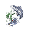
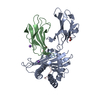
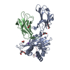
- PDB-8rbu: Crystal structure of HLA-A*11:01 in complex with SVLNDILARL, an 1... -
+ Open data

- Basic information
| Entry | Database: PDB / ID: 8rbu | ||||||
|---|---|---|---|---|---|---|---|
| Title | Crystal structure of HLA-A*11:01 in complex with SVLNDILARL, an 10-mer epitope from SARS-CoV-2 Spike (S975-984) | ||||||
 Components Components |
| ||||||
 Keywords Keywords | IMMUNE SYSTEM / human leukocyte antigen / major histocompatibility complex / HLA-A11 / HLA-A*11:01 / SARS-CoV-2 / Spike | ||||||
| Function / homology |  Function and homology information Function and homology informationantigen processing and presentation of peptide antigen via MHC class I / negative regulation of receptor binding / early endosome lumen / Nef mediated downregulation of MHC class I complex cell surface expression / DAP12 interactions / transferrin transport / cellular response to iron ion / lumenal side of endoplasmic reticulum membrane / Endosomal/Vacuolar pathway / Antigen Presentation: Folding, assembly and peptide loading of class I MHC ...antigen processing and presentation of peptide antigen via MHC class I / negative regulation of receptor binding / early endosome lumen / Nef mediated downregulation of MHC class I complex cell surface expression / DAP12 interactions / transferrin transport / cellular response to iron ion / lumenal side of endoplasmic reticulum membrane / Endosomal/Vacuolar pathway / Antigen Presentation: Folding, assembly and peptide loading of class I MHC / peptide antigen assembly with MHC class II protein complex / cellular response to iron(III) ion / MHC class II protein complex / negative regulation of forebrain neuron differentiation / antigen processing and presentation of exogenous protein antigen via MHC class Ib, TAP-dependent / ER to Golgi transport vesicle membrane / peptide antigen assembly with MHC class I protein complex / regulation of erythrocyte differentiation / regulation of iron ion transport / HFE-transferrin receptor complex / response to molecule of bacterial origin / MHC class I peptide loading complex / T cell mediated cytotoxicity / positive regulation of T cell cytokine production / antigen processing and presentation of endogenous peptide antigen via MHC class I / antigen processing and presentation of exogenous peptide antigen via MHC class II / positive regulation of immune response / MHC class I protein complex / positive regulation of T cell activation / peptide antigen binding / positive regulation of receptor-mediated endocytosis / negative regulation of neurogenesis / cellular response to nicotine / positive regulation of T cell mediated cytotoxicity / multicellular organismal-level iron ion homeostasis / specific granule lumen / phagocytic vesicle membrane / recycling endosome membrane / Immunoregulatory interactions between a Lymphoid and a non-Lymphoid cell / Interferon gamma signaling / negative regulation of epithelial cell proliferation / MHC class II protein complex binding / Modulation by Mtb of host immune system / late endosome membrane / sensory perception of smell / positive regulation of cellular senescence / tertiary granule lumen / DAP12 signaling / T cell differentiation in thymus / negative regulation of neuron projection development / ER-Phagosome pathway / protein refolding / early endosome membrane / symbiont-mediated disruption of host tissue / protein homotetramerization / Maturation of spike protein / Translation of Structural Proteins / Virion Assembly and Release / host cell surface / host extracellular space / viral translation / amyloid fibril formation / symbiont-mediated-mediated suppression of host tetherin activity / Induction of Cell-Cell Fusion / structural constituent of virion / intracellular iron ion homeostasis / entry receptor-mediated virion attachment to host cell / membrane fusion / Attachment and Entry / host cell endoplasmic reticulum-Golgi intermediate compartment membrane / learning or memory / positive regulation of viral entry into host cell / receptor-mediated virion attachment to host cell / host cell surface receptor binding / immune response / symbiont-mediated suppression of host innate immune response / receptor ligand activity / endocytosis involved in viral entry into host cell / endoplasmic reticulum lumen / Amyloid fiber formation / Golgi membrane / lysosomal membrane / fusion of virus membrane with host plasma membrane / external side of plasma membrane / focal adhesion / fusion of virus membrane with host endosome membrane / viral envelope / Neutrophil degranulation / symbiont entry into host cell / virion attachment to host cell / SARS-CoV-2 activates/modulates innate and adaptive immune responses / host cell plasma membrane / virion membrane / structural molecule activity / cell surface / endoplasmic reticulum / Golgi apparatus / protein homodimerization activity / extracellular space / extracellular exosome Similarity search - Function | ||||||
| Biological species |  Homo sapiens (human) Homo sapiens (human)  Severe acute respiratory syndrome coronavirus 2 Severe acute respiratory syndrome coronavirus 2 | ||||||
| Method |  X-RAY DIFFRACTION / SYNCHROTRON / MOLECULAR REPLACEMENT / Resolution: 2.7 Å X-RAY DIFFRACTION / SYNCHROTRON / MOLECULAR REPLACEMENT / Resolution: 2.7 Å | ||||||
 Authors Authors | Ahn, Y.M. / Maddumage, J.C. / Szeto, C. / Gras, S. | ||||||
| Funding support |  Australia, 1items Australia, 1items
| ||||||
 Citation Citation | Journal: Curr Res Struct Biol / Year: 2024 Title: The impact of SARS-CoV-2 spike mutation on peptide presentation is HLA allomorph-specific. Authors: Ahn, Y.M. / Maddumage, J.C. / Grant, E.J. / Chatzileontiadou, D.S.M. / Perera, W.W.J.G. / Baker, B.M. / Szeto, C. / Gras, S. | ||||||
| History |
|
- Structure visualization
| Structure viewer | Molecule: MolmilJmol/JSmol |
|---|
- Downloads & links
-Download
| PDBx/mmCIF format | 8rbu.cif.gz | 116.7 KB | Display | PDBx/mmCIF format |
|---|---|---|---|---|
| PDB format | pdb8rbu.ent.gz | 70.5 KB | Display | PDB format |
| PDBx/mmJSON format | 8rbu.json.gz | Tree view | PDBx/mmJSON format | |
| Others |  Other downloads Other downloads |
-Validation report
| Summary document | 8rbu_validation.pdf.gz | 3.7 MB | Display | wwPDB validaton report |
|---|---|---|---|---|
| Full document | 8rbu_full_validation.pdf.gz | 3.7 MB | Display | |
| Data in XML | 8rbu_validation.xml.gz | 15.5 KB | Display | |
| Data in CIF | 8rbu_validation.cif.gz | 20.5 KB | Display | |
| Arichive directory | https://data.pdbj.org/pub/pdb/validation_reports/rb/8rbuftp://data.pdbj.org/pub/pdb/validation_reports/rb/8rbu | HTTPS FTP |
-Related structure data


-Links
 PDBj
PDBj


- Assembly
| Deposited unit | 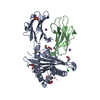
| ||||||||||||
|---|---|---|---|---|---|---|---|---|---|---|---|---|---|
| 1 |
| ||||||||||||
| Unit cell |
|
-Components
-Protein , 2 types, 2 molecules AB
| #1: Protein | Mass: 40985.727 Da / Num. of mol.: 1 Source method: isolated from a genetically manipulated source Source: (gene. exp.) Homo sapiens (human) / Gene: HLA-A, HLA, HLA-A11Production host:  References: UniProt: Q5S3G3 |
|---|---|
| #2: Protein | Mass: 11879.356 Da / Num. of mol.: 1 Source method: isolated from a genetically manipulated source Source: (gene. exp.) Homo sapiens (human) / Gene: B2M, CDABP0092, HDCMA22PProduction host: References: UniProt: P61769 |
-Protein/peptide , 1 types, 1 molecules C
| #3: Protein/peptide | Mass: 1114.317 Da / Num. of mol.: 1 / Source method: obtained synthetically Source: (synth.) Severe acute respiratory syndrome coronavirus 2References: UniProt: P0DTC2 |
|---|
-Non-polymers , 6 types, 31 molecules 


| #4: Chemical | ChemComp-PEG /  Mass: 106.120 Da / Num. of mol.: 1 / Source method: obtained synthetically / Formula: C4H10O3 / Feature type: SUBJECT OF INVESTIGATION Mass: 106.120 Da / Num. of mol.: 1 / Source method: obtained synthetically / Formula: C4H10O3 / Feature type: SUBJECT OF INVESTIGATION | ||||||||
|---|---|---|---|---|---|---|---|---|---|
| #5: Chemical | ChemComp-EDO /  Mass: 62.068 Da / Num. of mol.: 8 / Source method: obtained synthetically / Formula: C2H6O2 / Feature type: SUBJECT OF INVESTIGATION Mass: 62.068 Da / Num. of mol.: 8 / Source method: obtained synthetically / Formula: C2H6O2 / Feature type: SUBJECT OF INVESTIGATION#6: Chemical |  Mass: 35.453 Da / Num. of mol.: 2 / Source method: obtained synthetically / Formula: Cl / Feature type: SUBJECT OF INVESTIGATION Mass: 35.453 Da / Num. of mol.: 2 / Source method: obtained synthetically / Formula: Cl / Feature type: SUBJECT OF INVESTIGATION#7: Chemical |  Mass: 96.063 Da / Num. of mol.: 2 / Source method: obtained synthetically / Formula: SO4 / Feature type: SUBJECT OF INVESTIGATION Mass: 96.063 Da / Num. of mol.: 2 / Source method: obtained synthetically / Formula: SO4 / Feature type: SUBJECT OF INVESTIGATION#8: Chemical |  Mass: 22.990 Da / Num. of mol.: 2 / Source method: obtained synthetically / Formula: Na / Feature type: SUBJECT OF INVESTIGATION Mass: 22.990 Da / Num. of mol.: 2 / Source method: obtained synthetically / Formula: Na / Feature type: SUBJECT OF INVESTIGATION#9: Water | ChemComp-HOH / | Mass: 18.015 Da / Num. of mol.: 16 / Source method: isolated from a natural source / Formula: H2O |
-Details
| Has ligand of interest | Y |
|---|---|
| Has protein modification | Y |
-Experimental details
-Experiment
| Experiment | Method: X-RAY DIFFRACTION / Number of used crystals: 1 |
|---|
- Sample preparation
| Crystal | Density Matthews: 2.17 Å3/Da / Density % sol: 43.29 % |
|---|---|
| Crystal grow | Temperature: 293 K / Method: vapor diffusion, sitting drop / Details: 25% PEG3350, 0.2M NaCl and 0.1M Tris HCl pH8.5 |
-Data collection
| Diffraction | Mean temperature: 100 K / Serial crystal experiment: N |
|---|---|
| Diffraction source | Source: SYNCHROTRON / Site: Australian Synchrotron / Beamline: MX2 / Wavelength: 0.953732 Å |
| Detector | Type: DECTRIS EIGER X 16M / Detector: PIXEL / Date: Mar 23, 2022 |
| Radiation | Protocol: SINGLE WAVELENGTH / Monochromatic (M) / Laue (L): M / Scattering type: x-ray |
| Radiation wavelength | Wavelength: 0.953732 Å / Relative weight: 1 |
| Reflection | Resolution: 2.7→43.67 Å / Num. obs: 13337 / % possible obs: 100 % / Redundancy: 11 % / Biso Wilson estimate: 43.76 Å2 / CC1/2: 0.996 / Rmerge(I) obs: 0.175 / Rpim(I) all: 0.055 / Rrim(I) all: 0.184 / Net I/σ(I): 13 |
| Reflection shell | Resolution: 2.7→2.83 Å / Redundancy: 10.8 % / Rmerge(I) obs: 0.985 / Num. unique obs: 1734 / CC1/2: 0.773 / Rpim(I) all: 0.317 / Rrim(I) all: 1.036 / % possible all: 100 |
- Processing
| Software |
| |||||||||||||||||||||||||||||||||||
|---|---|---|---|---|---|---|---|---|---|---|---|---|---|---|---|---|---|---|---|---|---|---|---|---|---|---|---|---|---|---|---|---|---|---|---|---|
| Refinement | Method to determine structure: MOLECULAR REPLACEMENT / Resolution: 2.7→43.67 Å / SU ML: 0.3131 / Cross valid method: FREE R-VALUE / σ(F): 1.35 / Phase error: 26.3227 Stereochemistry target values: GeoStd + Monomer Library + CDL v1.2
| |||||||||||||||||||||||||||||||||||
| Solvent computation | Shrinkage radii: 0.9 Å / VDW probe radii: 1.1 Å / Solvent model: FLAT BULK SOLVENT MODEL | |||||||||||||||||||||||||||||||||||
| Displacement parameters | Biso mean: 43.9 Å2 | |||||||||||||||||||||||||||||||||||
| Refinement step | Cycle: LAST / Resolution: 2.7→43.67 Å
| |||||||||||||||||||||||||||||||||||
| Refine LS restraints |
| |||||||||||||||||||||||||||||||||||
| LS refinement shell |
|
