 Movie
Movie Controller


+ Open data

- Basic information
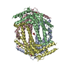
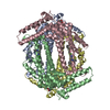
| Entry | Database: PDB / ID: 8r5r | ||||||
|---|---|---|---|---|---|---|---|
| Title | Structure of apo TDO with a bound inhibitor | ||||||
 Components Components | Tryptophan 2,3-dioxygenase | ||||||
 Keywords Keywords | OXIDOREDUCTASE / inhibitor / apo / heme | ||||||
| Function / homology |  Function and homology information Function and homology informationresponse to nitroglycerin / tryptophan 2,3-dioxygenase / : / L-tryptophan 2,3-dioxygenase activity / : / response to cortisol / Tryptophan catabolism / amino acid binding / oxygen binding / protein homotetramerization ...response to nitroglycerin / tryptophan 2,3-dioxygenase / : / L-tryptophan 2,3-dioxygenase activity / : / response to cortisol / Tryptophan catabolism / amino acid binding / oxygen binding / protein homotetramerization / response to ethanol / heme binding / metal ion binding / identical protein binding / cytosol Similarity search - Function | ||||||
| Biological species |  Homo sapiens (human) Homo sapiens (human) | ||||||
| Method |  X-RAY DIFFRACTION / SYNCHROTRON / MOLECULAR REPLACEMENT / Resolution: 3.078 Å X-RAY DIFFRACTION / SYNCHROTRON / MOLECULAR REPLACEMENT / Resolution: 3.078 Å | ||||||
 Authors Authors | Wicki, M. / Mac Sweeney, A. | ||||||
| Funding support | 1items
| ||||||
 Citation Citation | Journal: Sci Rep / Year: 2024 Title: Discovery and binding mode of small molecule inhibitors of the apo form of human TDO2. Authors: Lotz-Jenne, C. / Lange, R. / Cren, S. / Bourquin, G. / Goglia, L. / Kimmerlin, T. / Wicki, M. / Muller, M. / Artico, N. / Ackerknecht, S. / Pfaff, P. / Joesch, C. / Mac Sweeney, A. #1: Journal: Biorxiv / Year: 2024Title: Discovery and binding mode of a small molecule inhibitor of the apo form of human TDO2 Authors: Lotz-Jenne, C. / Lange, R. / Cren, S. / Bourquin, G. / Goglia, L. / Kimmerlin, T. / Wicki, M. / Mueller, M. / Artico, N. / Ackerknecht, S. / Joesch, C. / Mac Sweeney, A. | ||||||
| History |
|
- Structure visualization
| Structure viewer | Molecule: MolmilJmol/JSmol |
|---|
- Downloads & links
-Download
| PDBx/mmCIF format | 8r5r.cif.gz | 258.7 KB | Display | PDBx/mmCIF format |
|---|---|---|---|---|
| PDB format | pdb8r5r.ent.gz | 205.2 KB | Display | PDB format |
| PDBx/mmJSON format | 8r5r.json.gz | Tree view | PDBx/mmJSON format | |
| Others |  Other downloads Other downloads |
-Validation report
| Arichive directory | https://data.pdbj.org/pub/pdb/validation_reports/r5/8r5rftp://data.pdbj.org/pub/pdb/validation_reports/r5/8r5r | HTTPS FTP |
|---|
-Related structure data

-Links
 PDBj
PDBj- Assembly
| Deposited unit | 
| ||||||||
|---|---|---|---|---|---|---|---|---|---|
| 1 |
| ||||||||
| Unit cell |
|
-Components
| #1: Protein | Mass: 42878.027 Da / Num. of mol.: 4 Source method: isolated from a genetically manipulated source Source: (gene. exp.) Homo sapiens (human) / Gene: TDO2 / Production host:  #2: Chemical | ChemComp-Y5N / 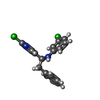  Mass: 343.250 Da / Num. of mol.: 4 / Source method: obtained synthetically / Formula: C19H16Cl2N2 / Feature type: SUBJECT OF INVESTIGATION Mass: 343.250 Da / Num. of mol.: 4 / Source method: obtained synthetically / Formula: C19H16Cl2N2 / Feature type: SUBJECT OF INVESTIGATION#3: Chemical | ChemComp-ZIQ /   Type: L-peptide linking / Mass: 218.252 Da / Num. of mol.: 4 / Source method: obtained synthetically / Formula: C12H14N2O2 Type: L-peptide linking / Mass: 218.252 Da / Num. of mol.: 4 / Source method: obtained synthetically / Formula: C12H14N2O2#4: Water | ChemComp-HOH / |  Mass: 18.015 Da / Num. of mol.: 7 / Source method: isolated from a natural source / Formula: H2O Mass: 18.015 Da / Num. of mol.: 7 / Source method: isolated from a natural source / Formula: H2OHas ligand of interest | Y | Has protein modification | N | |
|---|
-Experimental details
-Experiment
| Experiment | Method: X-RAY DIFFRACTION / Number of used crystals: 1 |
|---|
- Sample preparation
| Crystal | Density Matthews: 2.44 Å3/Da / Density % sol: 49.61 % |
|---|---|
| Crystal grow | Temperature: 293 K / Method: vapor diffusion, sitting drop / pH: 7.5 Details: 5.3 mg/ml (124 uM) protein containing 2 mM AMT and 1 mM inhibitor Reservoir solution: Morpheus II condition F8 5%(w/v) PEG 20K, 25%(w/v) 1,1,1-tris(hydroxymethyl)propane, 1%(w/v) NDSB 195,0. ...Details: 5.3 mg/ml (124 uM) protein containing 2 mM AMT and 1 mM inhibitor Reservoir solution: Morpheus II condition F8 5%(w/v) PEG 20K, 25%(w/v) 1,1,1-tris(hydroxymethyl)propane, 1%(w/v) NDSB 195,0.02M of each Monosaccharide II, 0.1M BES/TEA pH 7.5 |
-Data collection
| Diffraction | Mean temperature: 100 K / Serial crystal experiment: N |
|---|---|
| Diffraction source | Source: SYNCHROTRON / Site: ESRF  / Beamline: ID23-1 / Wavelength: 1 Å / Beamline: ID23-1 / Wavelength: 1 Å |
| Detector | Type: DECTRIS PILATUS 6M / Detector: PIXEL / Date: Oct 9, 2023 |
| Radiation | Protocol: SINGLE WAVELENGTH / Monochromatic (M) / Laue (L): M / Scattering type: x-ray |
| Radiation wavelength | Wavelength: 1 Å / Relative weight: 1 |
| Reflection | Resolution: 3.08→76.31 Å / Num. obs: 27716 / % possible obs: 95.2 % / Redundancy: 9.8 % / CC1/2: 0.998 / Rmerge(I) obs: 0.171 / Rpim(I) all: 0.056 / Net I/σ(I): 9.8 |
| Reflection shell | Resolution: 3.08→3.24 Å / Redundancy: 8.6 % / Mean I/σ(I) obs: 1.3 / Num. unique obs: 1386 / CC1/2: 0.536 / Rsym value: 0.648 / % possible all: 60.6 |
- Processing
| Software |
| ||||||||||||||||||||||||||||||||||||||||||||||||||||||||||||
|---|---|---|---|---|---|---|---|---|---|---|---|---|---|---|---|---|---|---|---|---|---|---|---|---|---|---|---|---|---|---|---|---|---|---|---|---|---|---|---|---|---|---|---|---|---|---|---|---|---|---|---|---|---|---|---|---|---|---|---|---|---|
| Refinement | Method to determine structure: MOLECULAR REPLACEMENT / Resolution: 3.078→50.83 Å / Cor.coef. Fo:Fc: 0.928 / Cor.coef. Fo:Fc free: 0.907 / Cross valid method: THROUGHOUT / SU Rfree Blow DPI: 0.474
| ||||||||||||||||||||||||||||||||||||||||||||||||||||||||||||
| Displacement parameters | Biso mean: 83.64 Å2
| ||||||||||||||||||||||||||||||||||||||||||||||||||||||||||||
| Refine analyze | Luzzati coordinate error obs: 0.46 Å | ||||||||||||||||||||||||||||||||||||||||||||||||||||||||||||
| Refinement step | Cycle: LAST / Resolution: 3.078→50.83 Å
| ||||||||||||||||||||||||||||||||||||||||||||||||||||||||||||
| Refine LS restraints |
| ||||||||||||||||||||||||||||||||||||||||||||||||||||||||||||
| LS refinement shell | Resolution: 3.08→3.18 Å
|
