 Movie
Movie Controller

[English] 日本語
 Yorodumi
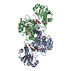
- PDB-8qzb: D-2-hydroxyacid dehydrogenase (D2HDH) from Haloferax mediterranei... -
+ Open data

- Basic information
| Entry | Database: PDB / ID: 8qzb | ||||||||||||||||||
|---|---|---|---|---|---|---|---|---|---|---|---|---|---|---|---|---|---|---|---|
| Title | D-2-hydroxyacid dehydrogenase (D2HDH) from Haloferax mediterranei in complex with 2-ketohexanoic acid, NAD+ and chloride (1.16 A resolution) | ||||||||||||||||||
 Components Components | D-2-hydroxyacid dehydrogenase | ||||||||||||||||||
 Keywords Keywords | OXIDOREDUCTASE / complex halophilic adaptation substrate specificity mechanism | ||||||||||||||||||
| Function / homology |  Function and homology information Function and homology informationNADH binding / carboxylic acid binding / carboxylic acid metabolic process / Oxidoreductases; Acting on the CH-OH group of donors; With NAD+ or NADP+ as acceptor / oxidoreductase activity, acting on the CH-OH group of donors, NAD or NADP as acceptor / NADPH binding Similarity search - Function | ||||||||||||||||||
| Biological species |  Haloferax mediterranei (archaea) Haloferax mediterranei (archaea) | ||||||||||||||||||
| Method |  X-RAY DIFFRACTION / SYNCHROTRON / MOLECULAR REPLACEMENT / Resolution: 1.16 Å X-RAY DIFFRACTION / SYNCHROTRON / MOLECULAR REPLACEMENT / Resolution: 1.16 Å | ||||||||||||||||||
 Authors Authors | Baker, P.J. / Barrett, J.R. / Dakhil, A.A.A.B. / Domenech, J. / Bisson, C. / Pramanpol, N. / Ferrer, J. / Rice, D.W. | ||||||||||||||||||
| Funding support |  United Kingdom, United Kingdom,  Spain, Spain,  Thailand, Libya, 5items Thailand, Libya, 5items
| ||||||||||||||||||
 Citation Citation | Journal: To Be Published Title: Ternary complexes of Haloferax mediterranei D-2-hydroxyacid dehydrogenase provide insights into halophilicity and the chiral specificity of its reaction mechanism. Authors: Domenech, J. / Pramanpol, N. / Bisson, C. / Sedelnikova, S.E. / Barrett, J.R. / Dakhil, A.A.A.B. / Abdelhameed, A.S. / Harding, S.E. / Rice, D.W. / Baker, P.J. / Ferrer, J. | ||||||||||||||||||
| History |
|
- Structure visualization
| Structure viewer | Molecule: MolmilJmol/JSmol |
|---|
- Downloads & links
-Download
| PDBx/mmCIF format | 8qzb.cif.gz | 739 KB | Display | PDBx/mmCIF format |
|---|---|---|---|---|
| PDB format | pdb8qzb.ent.gz | 464.1 KB | Display | PDB format |
| PDBx/mmJSON format | 8qzb.json.gz | Tree view | PDBx/mmJSON format | |
| Others |  Other downloads Other downloads |
-Validation report
| Summary document | 8qzb_validation.pdf.gz | 22.6 MB | Display | wwPDB validaton report |
|---|---|---|---|---|
| Full document | 8qzb_full_validation.pdf.gz | 22.7 MB | Display | |
| Data in XML | 8qzb_validation.xml.gz | 73.1 KB | Display | |
| Data in CIF | 8qzb_validation.cif.gz | 106.1 KB | Display | |
| Arichive directory | https://data.pdbj.org/pub/pdb/validation_reports/qz/8qzbftp://data.pdbj.org/pub/pdb/validation_reports/qz/8qzb | HTTPS FTP |
-Related structure data
| Related structure data | |
|---|---|
| Similar structure data |


-Links
 PDBj
PDBj- Assembly
| Deposited unit | 
| ||||||||
|---|---|---|---|---|---|---|---|---|---|
| 1 | 
| ||||||||
| 2 | 
| ||||||||
| Unit cell |
|
-Components
-Protein , 1 types, 4 molecules ABCD
| #1: Protein | Mass: 33367.992 Da / Num. of mol.: 4 Source method: isolated from a genetically manipulated source Source: (gene. exp.) Haloferax mediterranei (archaea) / Gene: ddh / Production host:  |
|---|
-Non-polymers , 7 types, 1666 molecules 
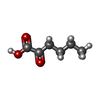


| #2: Chemical | ChemComp-NAD /  Mass: 663.425 Da / Num. of mol.: 4 / Source method: obtained synthetically / Formula: C21H27N7O14P2 / Feature type: SUBJECT OF INVESTIGATION / Comment: NAD*YM Mass: 663.425 Da / Num. of mol.: 4 / Source method: obtained synthetically / Formula: C21H27N7O14P2 / Feature type: SUBJECT OF INVESTIGATION / Comment: NAD*YM#3: Chemical | ChemComp-7N5 /  Mass: 130.142 Da / Num. of mol.: 4 / Source method: obtained synthetically / Formula: C6H10O3 / Feature type: SUBJECT OF INVESTIGATION Mass: 130.142 Da / Num. of mol.: 4 / Source method: obtained synthetically / Formula: C6H10O3 / Feature type: SUBJECT OF INVESTIGATION#4: Chemical | ChemComp-ACT /  Mass: 59.044 Da / Num. of mol.: 5 / Source method: obtained synthetically / Formula: C2H3O2 Mass: 59.044 Da / Num. of mol.: 5 / Source method: obtained synthetically / Formula: C2H3O2#5: Chemical | ChemComp-MG /  Mass: 24.305 Da / Num. of mol.: 18 / Source method: obtained synthetically / Formula: Mg / Feature type: SUBJECT OF INVESTIGATION Mass: 24.305 Da / Num. of mol.: 18 / Source method: obtained synthetically / Formula: Mg / Feature type: SUBJECT OF INVESTIGATION#6: Chemical | ChemComp-NA /  Mass: 22.990 Da / Num. of mol.: 12 / Source method: obtained synthetically / Formula: Na / Feature type: SUBJECT OF INVESTIGATION Mass: 22.990 Da / Num. of mol.: 12 / Source method: obtained synthetically / Formula: Na / Feature type: SUBJECT OF INVESTIGATION#7: Chemical | ChemComp-CL /  Mass: 35.453 Da / Num. of mol.: 4 / Source method: obtained synthetically / Formula: Cl / Feature type: SUBJECT OF INVESTIGATION Mass: 35.453 Da / Num. of mol.: 4 / Source method: obtained synthetically / Formula: Cl / Feature type: SUBJECT OF INVESTIGATION#8: Water | ChemComp-HOH / | Mass: 18.015 Da / Num. of mol.: 1619 / Source method: isolated from a natural source / Formula: H2O |
|---|
-Details
| Has ligand of interest | Y |
|---|---|
| Has protein modification | N |
-Experimental details
-Experiment
| Experiment | Method: X-RAY DIFFRACTION / Number of used crystals: 1 |
|---|
- Sample preparation
| Crystal | Density Matthews: 2.55 Å3/Da / Density % sol: 51.7 % / Description: blocks |
|---|---|
| Crystal grow | Temperature: 290 K / Method: vapor diffusion, hanging drop / pH: 8 Details: Protein buffer: 20mM Tris/HCl pH 8.0 2mM EDTA 1M NaCl 50mM 2-ketohexanoic acid 5mM NAD+ Crystallisation conditions: 0.1M Tris/HCl pH 8.0 0.5M Magnesium acetate 20% PEG 3350 |
-Data collection
| Diffraction | Mean temperature: 100 K / Serial crystal experiment: N |
|---|---|
| Diffraction source | Source: SYNCHROTRON / Site: Diamond / Beamline: I03 / Wavelength: 0.9801 Å |
| Detector | Type: DECTRIS EIGER2 XE 16M / Detector: PIXEL / Date: Feb 23, 2023 |
| Radiation | Protocol: SINGLE WAVELENGTH / Monochromatic (M) / Laue (L): M / Scattering type: x-ray |
| Radiation wavelength | Wavelength: 0.9801 Å / Relative weight: 1 |
| Reflection | Resolution: 1.16→46.633 Å / Num. obs: 427918 / % possible obs: 93.9 % / Redundancy: 3.6 % / CC1/2: 0.94 / Rpim(I) all: 0.042 / Net I/σ(I): 17.1 |
| Reflection shell | Resolution: 1.16→1.18 Å / Redundancy: 3.3 % / Num. unique obs: 18892 / CC1/2: 0.56 / Rpim(I) all: 0.5 / % possible all: 83.7 |
- Processing
| Software |
| |||||||||||||||||||||||||||||||||||||||||||||||||||||||||||||||||||||||||||||||||||||||||||||||||||||||||||||||||||||||||||||||||||||||||||||||||||||||||||||||||||||||||||||||||||||||||||||||||||||||||||||||||||||||||||||||||||||||
|---|---|---|---|---|---|---|---|---|---|---|---|---|---|---|---|---|---|---|---|---|---|---|---|---|---|---|---|---|---|---|---|---|---|---|---|---|---|---|---|---|---|---|---|---|---|---|---|---|---|---|---|---|---|---|---|---|---|---|---|---|---|---|---|---|---|---|---|---|---|---|---|---|---|---|---|---|---|---|---|---|---|---|---|---|---|---|---|---|---|---|---|---|---|---|---|---|---|---|---|---|---|---|---|---|---|---|---|---|---|---|---|---|---|---|---|---|---|---|---|---|---|---|---|---|---|---|---|---|---|---|---|---|---|---|---|---|---|---|---|---|---|---|---|---|---|---|---|---|---|---|---|---|---|---|---|---|---|---|---|---|---|---|---|---|---|---|---|---|---|---|---|---|---|---|---|---|---|---|---|---|---|---|---|---|---|---|---|---|---|---|---|---|---|---|---|---|---|---|---|---|---|---|---|---|---|---|---|---|---|---|---|---|---|---|---|---|---|---|---|---|---|---|---|---|---|---|---|---|---|---|---|---|
| Refinement | Method to determine structure: MOLECULAR REPLACEMENT / Resolution: 1.16→46.633 Å / Cor.coef. Fo:Fc: 0.982 / Cor.coef. Fo:Fc free: 0.975 / WRfactor Rfree: 0.15 / WRfactor Rwork: 0.122 / SU B: 1.258 / SU ML: 0.025 / Average fsc free: 0.9747 / Average fsc work: 0.9817 / Cross valid method: FREE R-VALUE / ESU R: 0.032 / ESU R Free: 0.033 Details: Hydrogens have been added in their riding positions
| |||||||||||||||||||||||||||||||||||||||||||||||||||||||||||||||||||||||||||||||||||||||||||||||||||||||||||||||||||||||||||||||||||||||||||||||||||||||||||||||||||||||||||||||||||||||||||||||||||||||||||||||||||||||||||||||||||||||
| Solvent computation | Ion probe radii: 0.8 Å / Shrinkage radii: 0.8 Å / VDW probe radii: 1.2 Å / Solvent model: MASK BULK SOLVENT | |||||||||||||||||||||||||||||||||||||||||||||||||||||||||||||||||||||||||||||||||||||||||||||||||||||||||||||||||||||||||||||||||||||||||||||||||||||||||||||||||||||||||||||||||||||||||||||||||||||||||||||||||||||||||||||||||||||||
| Displacement parameters | Biso mean: 14.538 Å2
| |||||||||||||||||||||||||||||||||||||||||||||||||||||||||||||||||||||||||||||||||||||||||||||||||||||||||||||||||||||||||||||||||||||||||||||||||||||||||||||||||||||||||||||||||||||||||||||||||||||||||||||||||||||||||||||||||||||||
| Refinement step | Cycle: LAST / Resolution: 1.16→46.633 Å
| |||||||||||||||||||||||||||||||||||||||||||||||||||||||||||||||||||||||||||||||||||||||||||||||||||||||||||||||||||||||||||||||||||||||||||||||||||||||||||||||||||||||||||||||||||||||||||||||||||||||||||||||||||||||||||||||||||||||
| Refine LS restraints |
| |||||||||||||||||||||||||||||||||||||||||||||||||||||||||||||||||||||||||||||||||||||||||||||||||||||||||||||||||||||||||||||||||||||||||||||||||||||||||||||||||||||||||||||||||||||||||||||||||||||||||||||||||||||||||||||||||||||||
| LS refinement shell | Refine-ID: X-RAY DIFFRACTION / Total num. of bins used: 20
|
