 Movie
Movie Controller


+ Open data

- Basic information
| Entry | Database: PDB / ID: 8qps | |||||||||||||||
|---|---|---|---|---|---|---|---|---|---|---|---|---|---|---|---|---|
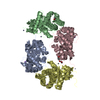
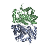
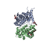
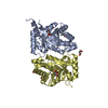
| Title | Deoxyribose-5-phosphate aldolase (DERA) from Geobacillus sp. | |||||||||||||||
 Components Components | Deoxyribose-phosphate aldolase | |||||||||||||||
 Keywords Keywords | LYASE / Aldolase / Autoinhibition / DERA / Thermophilic | |||||||||||||||
| Function / homology |  Function and homology information Function and homology informationdeoxyribose-phosphate aldolase / deoxyribose-phosphate aldolase activity / 2-deoxyribose 1-phosphate catabolic process / deoxyribonucleotide catabolic process / carbohydrate catabolic process / cytoplasm Similarity search - Function | |||||||||||||||
| Biological species |  Geobacillus sp. (bacteria) Geobacillus sp. (bacteria) | |||||||||||||||
| Method |  X-RAY DIFFRACTION / SYNCHROTRON / MOLECULAR REPLACEMENT / Resolution: 1.64 Å X-RAY DIFFRACTION / SYNCHROTRON / MOLECULAR REPLACEMENT / Resolution: 1.64 Å | |||||||||||||||
 Authors Authors | Paakkonen, J. / Hakulinen, N. / Rouvinen, J. | |||||||||||||||
| Funding support |  Finland, 4items Finland, 4items
| |||||||||||||||
 Citation Citation | Journal: To Be Published Title: Three-dimensional structure of deoxyribose phosphate aldolase (DERA) from thermophilic Geobacillus sp. Authors: Paakkonen, J. / Andberg, M. / Voutilainen, S. / Koivula, A. / Hakulinen, N. / Janis, J. / Rouvinen, J. | |||||||||||||||
| History |
|
- Structure visualization
| Structure viewer | Molecule: MolmilJmol/JSmol |
|---|
- Downloads & links
-Download
| PDBx/mmCIF format | 8qps.cif.gz | 122.2 KB | Display | PDBx/mmCIF format |
|---|---|---|---|---|
| PDB format | pdb8qps.ent.gz | 75.3 KB | Display | PDB format |
| PDBx/mmJSON format | 8qps.json.gz | Tree view | PDBx/mmJSON format | |
| Others |  Other downloads Other downloads |
-Validation report
| Summary document | 8qps_validation.pdf.gz | 429.1 KB | Display | wwPDB validaton report |
|---|---|---|---|---|
| Full document | 8qps_full_validation.pdf.gz | 431.7 KB | Display | |
| Data in XML | 8qps_validation.xml.gz | 22.5 KB | Display | |
| Data in CIF | 8qps_validation.cif.gz | 31.3 KB | Display | |
| Arichive directory | https://data.pdbj.org/pub/pdb/validation_reports/qp/8qpsftp://data.pdbj.org/pub/pdb/validation_reports/qp/8qps | HTTPS FTP |
-Related structure data
| Related structure data | |
|---|---|
| Similar structure data |

-Links
 PDBj
PDBj
- Assembly
| Deposited unit | 
| ||||||||||||
|---|---|---|---|---|---|---|---|---|---|---|---|---|---|
| 1 |
| ||||||||||||
| Unit cell |
|
-Components
| #1: Protein | Mass: 24262.604 Da / Num. of mol.: 2 Source method: isolated from a genetically manipulated source Source: (gene. exp.) Geobacillus sp. (bacteria) / Gene: deoC / Production host: #2: Water | ChemComp-HOH / |  Mass: 18.015 Da / Num. of mol.: 280 / Source method: isolated from a natural source / Formula: H2O Mass: 18.015 Da / Num. of mol.: 280 / Source method: isolated from a natural source / Formula: H2OHas protein modification | N | |
|---|
-Experimental details
-Experiment
| Experiment | Method: X-RAY DIFFRACTION / Number of used crystals: 1 |
|---|
- Sample preparation
| Crystal | Density Matthews: 1.9 Å3/Da / Density % sol: 35.4 % |
|---|---|
| Crystal grow | Temperature: 293 K / Method: vapor diffusion, hanging drop / pH: 8 / Details: 28% PEG 4000, 0.1 M Tris buffer |
-Data collection
| Diffraction | Mean temperature: 100 K / Serial crystal experiment: N |
|---|---|
| Diffraction source | Source: SYNCHROTRON / Site: Diamond  / Beamline: I24 / Wavelength: 0.9688 Å / Beamline: I24 / Wavelength: 0.9688 Å |
| Detector | Type: DECTRIS PILATUS3 6M / Detector: PIXEL / Date: Jul 22, 2019 |
| Radiation | Protocol: SINGLE WAVELENGTH / Monochromatic (M) / Laue (L): M / Scattering type: x-ray |
| Radiation wavelength | Wavelength: 0.9688 Å / Relative weight: 1 |
| Reflection | Resolution: 1.64→34.68 Å / Num. obs: 42879 / % possible obs: 98.4 % / Redundancy: 2.56 % / Biso Wilson estimate: 28.2 Å2 / CC1/2: 0.993 / Rmerge(I) obs: 0.057 / Rpim(I) all: 0.043 / Rrim(I) all: 0.072 / Net I/σ(I): 10.1 |
| Reflection shell | Resolution: 1.64→1.7 Å / Redundancy: 2.58 % / Rmerge(I) obs: 0.684 / Mean I/σ(I) obs: 1.3 / Num. unique obs: 4272 / CC1/2: 0.503 / Rpim(I) all: 0.496 / Rrim(I) all: 0.854 / % possible all: 98.3 |
- Processing
| Software |
| |||||||||||||||||||||||||||||||||||||||||||||||||||||||||||||||||||||||||||||||||||||||||||||||||||||||||
|---|---|---|---|---|---|---|---|---|---|---|---|---|---|---|---|---|---|---|---|---|---|---|---|---|---|---|---|---|---|---|---|---|---|---|---|---|---|---|---|---|---|---|---|---|---|---|---|---|---|---|---|---|---|---|---|---|---|---|---|---|---|---|---|---|---|---|---|---|---|---|---|---|---|---|---|---|---|---|---|---|---|---|---|---|---|---|---|---|---|---|---|---|---|---|---|---|---|---|---|---|---|---|---|---|---|---|
| Refinement | Method to determine structure: MOLECULAR REPLACEMENT / Resolution: 1.64→34.67 Å / SU ML: 0.2453 / Cross valid method: FREE R-VALUE / σ(F): 1.96 / Phase error: 31.6539 Stereochemistry target values: GeoStd + Monomer Library + CDL v1.2
| |||||||||||||||||||||||||||||||||||||||||||||||||||||||||||||||||||||||||||||||||||||||||||||||||||||||||
| Solvent computation | Shrinkage radii: 0.9 Å / VDW probe radii: 1.11 Å / Solvent model: FLAT BULK SOLVENT MODEL | |||||||||||||||||||||||||||||||||||||||||||||||||||||||||||||||||||||||||||||||||||||||||||||||||||||||||
| Displacement parameters | Biso mean: 31.49 Å2 | |||||||||||||||||||||||||||||||||||||||||||||||||||||||||||||||||||||||||||||||||||||||||||||||||||||||||
| Refinement step | Cycle: LAST / Resolution: 1.64→34.67 Å
| |||||||||||||||||||||||||||||||||||||||||||||||||||||||||||||||||||||||||||||||||||||||||||||||||||||||||
| Refine LS restraints |
| |||||||||||||||||||||||||||||||||||||||||||||||||||||||||||||||||||||||||||||||||||||||||||||||||||||||||
| LS refinement shell |
|
