 Movie
Movie Controller


+ Open data

- Basic information
| Entry | Database: PDB / ID: 8qou | ||||||
|---|---|---|---|---|---|---|---|
| Title | Reactive intermediate deaminase A mutant - R107K | ||||||
 Components Components | 2-iminobutanoate/2-iminopropanoate deaminase | ||||||
 Keywords Keywords | UNKNOWN FUNCTION / Enzyme / deaminase | ||||||
| Function / homology |  Function and homology information Function and homology information2-iminobutanoate/2-iminopropanoate deaminase / 2-iminobutanoate deaminase activity / 2-iminopropanoate deaminase activity / mRNA destabilization / mRNA catabolic process / lipid metabolic process / peroxisome / RNA endonuclease activity producing 3'-phosphomonoesters, hydrolytic mechanism / Hydrolases; Acting on ester bonds / negative regulation of translation ...2-iminobutanoate/2-iminopropanoate deaminase / 2-iminobutanoate deaminase activity / 2-iminopropanoate deaminase activity / mRNA destabilization / mRNA catabolic process / lipid metabolic process / peroxisome / RNA endonuclease activity producing 3'-phosphomonoesters, hydrolytic mechanism / Hydrolases; Acting on ester bonds / negative regulation of translation / mitochondrial matrix / mRNA binding / nucleus / cytosol Similarity search - Function | ||||||
| Biological species |  | ||||||
| Method |  X-RAY DIFFRACTION / SYNCHROTRON / MOLECULAR REPLACEMENT / Resolution: 1.4 Å X-RAY DIFFRACTION / SYNCHROTRON / MOLECULAR REPLACEMENT / Resolution: 1.4 Å | ||||||
 Authors Authors | Rizzi, G. / Visentin, C. / Di Pisa, F. / Ricagno, S. | ||||||
| Funding support |  Italy, 1items Italy, 1items
| ||||||
 Citation Citation | Journal: Protein Sci. / Year: 2024 Title: Site-directed mutagenesis reveals the interplay between stability, structure, and enzymatic activity in RidA from Capra hircus. Authors: Rizzi, G. / Digiovanni, S. / Degani, G. / Barbiroli, A. / Di Pisa, F. / Popolo, L. / Visentin, C. / Vanoni, M.A. / Ricagno, S. | ||||||
| History |
|
- Structure visualization
| Structure viewer | Molecule: MolmilJmol/JSmol |
|---|
- Downloads & links
-Download
| PDBx/mmCIF format | 8qou.cif.gz | 131.3 KB | Display | PDBx/mmCIF format |
|---|---|---|---|---|
| PDB format | pdb8qou.ent.gz | 101.9 KB | Display | PDB format |
| PDBx/mmJSON format | 8qou.json.gz | Tree view | PDBx/mmJSON format | |
| Others |  Other downloads Other downloads |
-Validation report
| Summary document | 8qou_validation.pdf.gz | 1.9 MB | Display | wwPDB validaton report |
|---|---|---|---|---|
| Full document | 8qou_full_validation.pdf.gz | 1.9 MB | Display | |
| Data in XML | 8qou_validation.xml.gz | 15.9 KB | Display | |
| Data in CIF | 8qou_validation.cif.gz | 23.8 KB | Display | |
| Arichive directory | https://data.pdbj.org/pub/pdb/validation_reports/qo/8qouftp://data.pdbj.org/pub/pdb/validation_reports/qo/8qou | HTTPS FTP |
-Related structure data
| Related structure data | 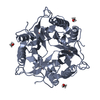 8q3hC  8qokC 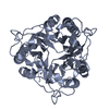 8qomC 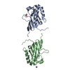 8qopC 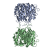 8qoqC 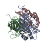 8qosC 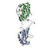 8qovC C: citing same article ( |
|---|---|
| Similar structure data |
-Links
 PDBj
PDBj
- Assembly
| Deposited unit | 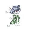
| |||||||||||||||||||||||||||
|---|---|---|---|---|---|---|---|---|---|---|---|---|---|---|---|---|---|---|---|---|---|---|---|---|---|---|---|---|
| 1 | 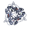
| |||||||||||||||||||||||||||
| 2 | 
| |||||||||||||||||||||||||||
| Unit cell |
| |||||||||||||||||||||||||||
| Components on special symmetry positions |
|
-Components
| #1: Protein | Mass: 14563.620 Da / Num. of mol.: 2 Source method: isolated from a genetically manipulated source Source: (gene. exp.)  References: UniProt: P80601, 2-iminobutanoate/2-iminopropanoate deaminase, Hydrolases; Acting on ester bonds #2: Chemical | ChemComp-PEG / |   Mass: 106.120 Da / Num. of mol.: 1 / Source method: obtained synthetically / Formula: C4H10O3 / Feature type: SUBJECT OF INVESTIGATION Mass: 106.120 Da / Num. of mol.: 1 / Source method: obtained synthetically / Formula: C4H10O3 / Feature type: SUBJECT OF INVESTIGATION#3: Chemical | ChemComp-EDO / |   Mass: 62.068 Da / Num. of mol.: 1 / Source method: obtained synthetically / Formula: C2H6O2 / Feature type: SUBJECT OF INVESTIGATION Mass: 62.068 Da / Num. of mol.: 1 / Source method: obtained synthetically / Formula: C2H6O2 / Feature type: SUBJECT OF INVESTIGATION#4: Chemical |   Mass: 58.693 Da / Num. of mol.: 2 / Source method: obtained synthetically / Formula: Ni / Feature type: SUBJECT OF INVESTIGATION Mass: 58.693 Da / Num. of mol.: 2 / Source method: obtained synthetically / Formula: Ni / Feature type: SUBJECT OF INVESTIGATION#5: Water | ChemComp-HOH / |  Mass: 18.015 Da / Num. of mol.: 340 / Source method: isolated from a natural source / Formula: H2O Mass: 18.015 Da / Num. of mol.: 340 / Source method: isolated from a natural source / Formula: H2OHas ligand of interest | Y | Has protein modification | N | |
|---|
-Experimental details
-Experiment
| Experiment | Method: X-RAY DIFFRACTION / Number of used crystals: 1 |
|---|
- Sample preparation
| Crystal | Density Matthews: 2.13 Å3/Da / Density % sol: 42.28 % |
|---|---|
| Crystal grow | Temperature: 298 K / Method: vapor diffusion, sitting drop / pH: 6.5 Details: 0.2 M Ammonium sulfate, 0.1 M Sodium cacodylate trihydrate, pH 6.5, 30% w/v polyethylene glycol 8000 |
-Data collection
| Diffraction | Mean temperature: 100 K / Serial crystal experiment: N |
|---|---|
| Diffraction source | Source: SYNCHROTRON / Site: ESRF  / Beamline: ID23-2 / Wavelength: 0.873 Å / Beamline: ID23-2 / Wavelength: 0.873 Å |
| Detector | Type: DECTRIS EIGER X 9M / Detector: PIXEL / Date: Nov 7, 2020 |
| Radiation | Protocol: SINGLE WAVELENGTH / Monochromatic (M) / Laue (L): M / Scattering type: x-ray |
| Radiation wavelength | Wavelength: 0.873 Å / Relative weight: 1 |
| Reflection | Resolution: 1.4→45.24 Å / Num. obs: 48575 / % possible obs: 99.7 % / Redundancy: 1.9 % / CC1/2: 0.939 / Net I/σ(I): 17.4 |
| Reflection shell | Resolution: 1.4→1.42 Å / Mean I/σ(I) obs: 4.7 / Num. unique obs: 2408 / CC1/2: 0.588 |
- Processing
| Software |
| ||||||||||||||||||||||||||||||||||||||||||||||||||||||||||||||||||||||||||||||||||||||||||||||||||||||||||||||||||||||||||||||||||||||||||||||||||||||||||||||||||||||||||||||||||||||
|---|---|---|---|---|---|---|---|---|---|---|---|---|---|---|---|---|---|---|---|---|---|---|---|---|---|---|---|---|---|---|---|---|---|---|---|---|---|---|---|---|---|---|---|---|---|---|---|---|---|---|---|---|---|---|---|---|---|---|---|---|---|---|---|---|---|---|---|---|---|---|---|---|---|---|---|---|---|---|---|---|---|---|---|---|---|---|---|---|---|---|---|---|---|---|---|---|---|---|---|---|---|---|---|---|---|---|---|---|---|---|---|---|---|---|---|---|---|---|---|---|---|---|---|---|---|---|---|---|---|---|---|---|---|---|---|---|---|---|---|---|---|---|---|---|---|---|---|---|---|---|---|---|---|---|---|---|---|---|---|---|---|---|---|---|---|---|---|---|---|---|---|---|---|---|---|---|---|---|---|---|---|---|---|
| Refinement | Method to determine structure: MOLECULAR REPLACEMENT / Resolution: 1.4→40.5 Å / Cor.coef. Fo:Fc: 0.954 / Cor.coef. Fo:Fc free: 0.953 / SU B: 1.512 / SU ML: 0.029 / Cross valid method: THROUGHOUT / ESU R: 0.067 / ESU R Free: 0.056 / Stereochemistry target values: MAXIMUM LIKELIHOOD / Details: HYDROGENS HAVE BEEN ADDED IN THE RIDING POSITIONS
| ||||||||||||||||||||||||||||||||||||||||||||||||||||||||||||||||||||||||||||||||||||||||||||||||||||||||||||||||||||||||||||||||||||||||||||||||||||||||||||||||||||||||||||||||||||||
| Solvent computation | Ion probe radii: 0.8 Å / Shrinkage radii: 0.8 Å / VDW probe radii: 1.2 Å / Solvent model: MASK | ||||||||||||||||||||||||||||||||||||||||||||||||||||||||||||||||||||||||||||||||||||||||||||||||||||||||||||||||||||||||||||||||||||||||||||||||||||||||||||||||||||||||||||||||||||||
| Displacement parameters | Biso mean: 10.93 Å2
| ||||||||||||||||||||||||||||||||||||||||||||||||||||||||||||||||||||||||||||||||||||||||||||||||||||||||||||||||||||||||||||||||||||||||||||||||||||||||||||||||||||||||||||||||||||||
| Refinement step | Cycle: 1 / Resolution: 1.4→40.5 Å
| ||||||||||||||||||||||||||||||||||||||||||||||||||||||||||||||||||||||||||||||||||||||||||||||||||||||||||||||||||||||||||||||||||||||||||||||||||||||||||||||||||||||||||||||||||||||
| Refine LS restraints |
|