Movie
Movie Controller

[English] 日本語
 Yorodumi
- PDB-8qot: Structure of the mu opioid receptor bound to the antagonist nanob... -
+ Open data

- Basic information
| Entry | Database: PDB / ID: 8qot | ||||||||||||
|---|---|---|---|---|---|---|---|---|---|---|---|---|---|
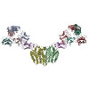
| Title | Structure of the mu opioid receptor bound to the antagonist nanobody NbE | ||||||||||||
 Components Components |
| ||||||||||||
 Keywords Keywords | MEMBRANE PROTEIN / opioid receptor / nanobody / antagonist / NabFab / MOR | ||||||||||||
| Function / homology |  Function and homology information Function and homology informationOpioid Signalling / spine apparatus / positive regulation of appetite / sperm ejaculation / Peptide ligand-binding receptors / G-protein activation / beta-endorphin receptor activity / morphine receptor activity / negative regulation of Wnt protein secretion / adenylate cyclase-inhibiting opioid receptor signaling pathway ...Opioid Signalling / spine apparatus / positive regulation of appetite / sperm ejaculation / Peptide ligand-binding receptors / G-protein activation / beta-endorphin receptor activity / morphine receptor activity / negative regulation of Wnt protein secretion / adenylate cyclase-inhibiting opioid receptor signaling pathway / negative regulation of luteinizing hormone secretion / G protein-coupled opioid receptor activity / regulation of cellular response to stress / G protein-coupled opioid receptor signaling pathway / G alpha (i) signalling events / negative regulation of nitric oxide biosynthetic process / negative regulation of adenylate cyclase-activating G protein-coupled receptor signaling pathway / adenylate cyclase-inhibiting G protein-coupled acetylcholine receptor signaling pathway / behavioral response to ethanol / regulation of NMDA receptor activity / neuropeptide binding / positive regulation of neurogenesis / eating behavior / negative regulation of cytosolic calcium ion concentration / transmission of nerve impulse / G-protein alpha-subunit binding / social behavior / voltage-gated calcium channel activity / T-tubule / sensory perception of pain / positive regulation of gluconeogenesis / dendrite membrane / dendrite cytoplasm / presynaptic modulation of chemical synaptic transmission / sarcoplasmic reticulum / neuropeptide signaling pathway / excitatory postsynaptic potential / locomotory behavior / sarcolemma / GABA-ergic synapse / G protein-coupled receptor activity / adenylate cyclase-inhibiting G protein-coupled receptor signaling pathway / adenylate cyclase-activating dopamine receptor signaling pathway / G-protein beta-subunit binding / presynaptic membrane / phospholipase C-activating G protein-coupled receptor signaling pathway / response to ethanol / perikaryon / positive regulation of ERK1 and ERK2 cascade / postsynaptic membrane / endosome / neuron projection / G protein-coupled receptor signaling pathway / protein domain specific binding / axon / focal adhesion / dendrite / mitochondrion / membrane / plasma membrane Similarity search - Function | ||||||||||||
| Biological species |  synthetic construct (others) | ||||||||||||
| Method | ELECTRON MICROSCOPY / single particle reconstruction / cryo EM / Resolution: 3.2 Å | ||||||||||||
 Authors Authors | Yu, J. / Kumar, A. / Zhang, X. / Martin, C. / Raia, P. / Manglik, A. / Ballet, S. / Boland, A. / Stoeber, M. | ||||||||||||
| Funding support |  Switzerland, 3items Switzerland, 3items
| ||||||||||||
 Citation Citation | Journal: Nat Commun / Year: 2024 Title: Structural basis of μ-opioid receptor targeting by a nanobody antagonist. Authors: Jun Yu / Amit Kumar / Xuefeng Zhang / Charlotte Martin / Kevin Van Holsbeeck / Pierre Raia / Antoine Koehl / Toon Laeremans / Jan Steyaert / Aashish Manglik / Steven Ballet / Andreas Boland / Miriam Stoeber /   Abstract: The μ-opioid receptor (μOR), a prototypical G protein-coupled receptor (GPCR), is the target of opioid analgesics such as morphine and fentanyl. Due to the severe side effects of current opioid ...The μ-opioid receptor (μOR), a prototypical G protein-coupled receptor (GPCR), is the target of opioid analgesics such as morphine and fentanyl. Due to the severe side effects of current opioid drugs, there is considerable interest in developing novel modulators of μOR function. Most GPCR ligands today are small molecules, however biologics, including antibodies and nanobodies, represent alternative therapeutics with clear advantages such as affinity and target selectivity. Here, we describe the nanobody NbE, which selectively binds to the μOR and acts as an antagonist. We functionally characterize NbE as an extracellular and genetically encoded μOR ligand and uncover the molecular basis for μOR antagonism by determining the cryo-EM structure of the NbE-μOR complex. NbE displays a unique ligand binding mode and achieves μOR selectivity by interactions with the orthosteric pocket and extracellular receptor loops. Based on a β-hairpin loop formed by NbE that deeply protrudes into the μOR, we design linear and cyclic peptide analogs that recapitulate NbE's antagonism. The work illustrates the potential of nanobodies to uniquely engage with GPCRs and describes lower molecular weight μOR ligands that can serve as a basis for therapeutic developments. #1: Journal: bioRxiv / Year: 2023 Title: Structural Basis of μ-Opioid Receptor-Targeting by a Nanobody Antagonist. Authors: Jun Yu / Amit Kumar / Xuefeng Zhang / Charlotte Martin / Pierre Raia / Antoine Koehl / Toon Laeremans / Jan Steyaert / Aashish Manglik / Steven Ballet / Andreas Boland / Miriam Stoeber / Abstract: The μ-opioid receptor (μOR), a prototypical member of the G protein-coupled receptor (GPCR) family, is the molecular target of opioid analgesics such as morphine and fentanyl. Due to the ...The μ-opioid receptor (μOR), a prototypical member of the G protein-coupled receptor (GPCR) family, is the molecular target of opioid analgesics such as morphine and fentanyl. Due to the limitations and severe side effects of currently available opioid drugs, there is considerable interest in developing novel modulators of μOR function. Most GPCR ligands today are small molecules, however biologics, including antibodies and nanobodies, are emerging as alternative therapeutics with clear advantages such as affinity and target selectivity. Here, we describe the nanobody NbE, which selectively binds to the μOR and acts as an antagonist. We functionally characterize NbE as an extracellular and genetically encoded μOR ligand and uncover the molecular basis for μOR antagonism by solving the cryo-EM structure of the NbE-μOR complex. NbE displays a unique ligand binding mode and achieves μOR selectivity by interactions with the orthosteric pocket and extracellular receptor loops. Based on a β-hairpin loop formed by NbE that deeply inserts into the μOR and centers most binding contacts, we design short peptide analogues that retain μOR antagonism. The work illustrates the potential of nanobodies to uniquely engage with GPCRs and describes novel μOR ligands that can serve as a basis for therapeutic developments. | ||||||||||||
| History |
|
- Structure visualization
| Structure viewer | Molecule: MolmilJmol/JSmol |
|---|
- Downloads & links
-Download
| PDBx/mmCIF format | 8qot.cif.gz | 190 KB | Display | PDBx/mmCIF format |
|---|---|---|---|---|
| PDB format | pdb8qot.ent.gz | 141 KB | Display | PDB format |
| PDBx/mmJSON format | 8qot.json.gz | Tree view | PDBx/mmJSON format | |
| Others |  Other downloads Other downloads |
-Validation report
| Arichive directory | https://data.pdbj.org/pub/pdb/validation_reports/qo/8qotftp://data.pdbj.org/pub/pdb/validation_reports/qo/8qot | HTTPS FTP |
|---|
-Related structure data
| Related structure data |  18541MC  8v8kC M: map data used to model this data C: citing same article ( |
|---|---|
| Similar structure data |
-Links
 PDBj
PDBj


- Assembly
| Deposited unit | 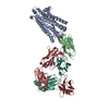
|
|---|---|
| 1 |
|
-Components
| #1: Protein | Mass: 53994.215 Da / Num. of mol.: 1 Source method: isolated from a genetically manipulated source Details: MOR-2xSTREP-8xHIS / Source: (gene. exp.)   Spodoptera frugiperda (fall armyworm) / References: UniProt: P42866 Spodoptera frugiperda (fall armyworm) / References: UniProt: P42866 |
|---|---|
| #2: Antibody | Mass: 18458.662 Da / Num. of mol.: 1 Source method: isolated from a genetically manipulated source Details: synthesized nanobody NbE-6xHis / Source: (gene. exp.)  |
| #3: Antibody | Mass: 28220.525 Da / Num. of mol.: 1 Source method: isolated from a genetically manipulated source Details: Signal sequence-NabFab HC / Source: (gene. exp.) synthetic construct (others) / Production host: |
| #4: Antibody | Mass: 25794.859 Da / Num. of mol.: 1 Source method: isolated from a genetically manipulated source Source: (gene. exp.) synthetic construct (others) / Production host: |
| #5: Antibody | Mass: 17414.383 Da / Num. of mol.: 1 Source method: isolated from a genetically manipulated source Details: Signal sequence-6xHis Anti-Fab nanobody / Source: (gene. exp.) |
| Has protein modification | Y |
-Experimental details
-Experiment
| Experiment | Method: ELECTRON MICROSCOPY |
|---|---|
| EM experiment | Aggregation state: PARTICLE / 3D reconstruction method: single particle reconstruction |
- Sample preparation
| Component | Name: MOR-NbE-NabFab-AntiFab complex / Type: COMPLEX Details: Mu-opioid receptor bound to a nanobody antagonist (NbE), and NabFab module used as fiducial marker. Entity ID: all / Source: MULTIPLE SOURCES | |||||||||||||||||||||||||
|---|---|---|---|---|---|---|---|---|---|---|---|---|---|---|---|---|---|---|---|---|---|---|---|---|---|---|
| Molecular weight | Value: 0.12 MDa | |||||||||||||||||||||||||
| Source (natural) | Organism: | |||||||||||||||||||||||||
| Source (recombinant) | Organism: Spodoptera frugiperda (fall armyworm) | |||||||||||||||||||||||||
| Buffer solution | pH: 7.5 | |||||||||||||||||||||||||
| Buffer component |
| |||||||||||||||||||||||||
| Specimen | Conc.: 2.8 mg/ml / Embedding applied: NO / Shadowing applied: NO / Staining applied: NO / Vitrification applied: YES Details: MOR receptor was expressed and purified in insect cells in isolation. NbE was expressed and purified in E. coli. NabFab module was expressed and purified in E. coli. All purified components ...Details: MOR receptor was expressed and purified in insect cells in isolation. NbE was expressed and purified in E. coli. NabFab module was expressed and purified in E. coli. All purified components were incubated and formed readily a stable complex. Final complex was purified over size exclusion chromatography. | |||||||||||||||||||||||||
| Specimen support | Grid material: GOLD / Grid mesh size: 300 divisions/in. / Grid type: UltrAuFoil R1.2/1.3 | |||||||||||||||||||||||||
| Vitrification | Instrument: LEICA EM GP / Cryogen name: ETHANE / Humidity: 95 % / Chamber temperature: 288 K |
- Electron microscopy imaging
| Experimental equipment |  Model: Talos Arctica / Image courtesy: FEI Company |
|---|---|
| Microscopy | Model: FEI TALOS ARCTICA |
| Electron gun | Electron source:  FIELD EMISSION GUN / Accelerating voltage: 200 kV / Illumination mode: FLOOD BEAM FIELD EMISSION GUN / Accelerating voltage: 200 kV / Illumination mode: FLOOD BEAM |
| Electron lens | Mode: BRIGHT FIELD / Nominal magnification: 150000 X / Nominal defocus max: 2000 nm / Nominal defocus min: 600 nm / Cs: 2.7 mm / C2 aperture diameter: 70 µm / Alignment procedure: COMA FREE |
| Specimen holder | Cryogen: NITROGEN / Specimen holder model: FEI TITAN KRIOS AUTOGRID HOLDER / Temperature (max): 95 K / Temperature (min): 90 K |
| Image recording | Average exposure time: 52 sec. / Electron dose: 40 e/Å2 / Detector mode: COUNTING / Film or detector model: FEI FALCON III (4k x 4k) / Num. of grids imaged: 4 / Num. of real images: 5938 |
| Image scans | Width: 4096 / Height: 4096 |
- Processing
| EM software |
| ||||||||||||||||||||||||||||||||||||||||
|---|---|---|---|---|---|---|---|---|---|---|---|---|---|---|---|---|---|---|---|---|---|---|---|---|---|---|---|---|---|---|---|---|---|---|---|---|---|---|---|---|---|
| CTF correction | Type: NONE | ||||||||||||||||||||||||||||||||||||||||
| Particle selection | Num. of particles selected: 6063911 | ||||||||||||||||||||||||||||||||||||||||
| 3D reconstruction | Resolution: 3.2 Å / Resolution method: FSC 0.143 CUT-OFF / Num. of particles: 445766 / Num. of class averages: 1 / Symmetry type: POINT | ||||||||||||||||||||||||||||||||||||||||
| Atomic model building | B value: 68 / Protocol: AB INITIO MODEL / Space: REAL | ||||||||||||||||||||||||||||||||||||||||
| Atomic model building | 3D fitting-ID: 1 / Source name: PDB / Type: experimental model
| ||||||||||||||||||||||||||||||||||||||||
| Refinement | Cross valid method: NONE |