[English] 日本語
Yorodumi
- PDB-4dkl: Crystal structure of the mu-opioid receptor bound to a morphinan ... -

+
Open data


ID or keywords:

Loading...

-
Basic information

Entry
Database: PDB / ID: 4dkl
TitleCrystal structure of the mu-opioid receptor bound to a morphinan antagonist
ComponentsMu-type opioid receptor, lysozyme chimera
KeywordsSIGNALING PROTEIN/ANTAGONIST / G-protein coupled receptor / 7 transmembrane receptor / SIGNALING PROTEIN-ANTAGONIST complex
Function / homology
Function and homology information


Opioid Signalling / spine apparatus / beta-endorphin receptor activity / morphine receptor activity / negative regulation of Wnt protein secretion / negative regulation of adenylate cyclase-activating G protein-coupled receptor signaling pathway / Peptide ligand-binding receptors / adenylate cyclase-inhibiting opioid receptor signaling pathway / positive regulation of appetite / G-protein activation ...Opioid Signalling / spine apparatus / beta-endorphin receptor activity / morphine receptor activity / negative regulation of Wnt protein secretion / negative regulation of adenylate cyclase-activating G protein-coupled receptor signaling pathway / Peptide ligand-binding receptors / adenylate cyclase-inhibiting opioid receptor signaling pathway / positive regulation of appetite / G-protein activation / G protein-coupled opioid receptor activity / filamin binding / G protein-coupled opioid receptor signaling pathway / regulation of cellular response to stress / G alpha (i) signalling events / adenylate cyclase-inhibiting G protein-coupled acetylcholine receptor signaling pathway / behavioral response to ethanol / negative regulation of nitric oxide biosynthetic process / neuropeptide binding / negative regulation of cAMP-mediated signaling / positive regulation of neurogenesis / eating behavior / negative regulation of cytosolic calcium ion concentration / transmission of nerve impulse / regulation of NMDA receptor activity / GABA-ergic synapse / neuropeptide signaling pathway / G-protein alpha-subunit binding / voltage-gated calcium channel activity / positive regulation of cAMP-mediated signaling / sensory perception of pain / viral release from host cell by cytolysis / dendrite membrane / adenylate cyclase-inhibiting G protein-coupled receptor signaling pathway / excitatory postsynaptic potential / presynaptic modulation of chemical synaptic transmission / dendrite cytoplasm / peptidoglycan catabolic process / locomotory behavior / G protein-coupled receptor activity / adenylate cyclase-activating dopamine receptor signaling pathway / G-protein beta-subunit binding / positive regulation of nitric oxide biosynthetic process / cell wall macromolecule catabolic process / presynaptic membrane / lysozyme / lysozyme activity / phospholipase C-activating G protein-coupled receptor signaling pathway / positive regulation of cytosolic calcium ion concentration / perikaryon / postsynaptic membrane / response to ethanol / host cell cytoplasm / positive regulation of ERK1 and ERK2 cascade / endosome / neuron projection / defense response to bacterium / G protein-coupled receptor signaling pathway / axon / protein domain specific binding / focal adhesion / dendrite / membrane / plasma membrane
Similarity search - Function
Mu opioid receptor / Opioid receptor / Rhopdopsin 7-helix transmembrane proteins / Rhodopsin 7-helix transmembrane proteins / Lysozyme - #40 / Endolysin T4 type / T4-type lysozyme / Glycoside hydrolase, family 24 / Lysozyme domain superfamily / Phage lysozyme ...Mu opioid receptor / Opioid receptor / Rhopdopsin 7-helix transmembrane proteins / Rhodopsin 7-helix transmembrane proteins / Lysozyme - #40 / Endolysin T4 type / T4-type lysozyme / Glycoside hydrolase, family 24 / Lysozyme domain superfamily / Phage lysozyme / Lysozyme / G-protein coupled receptors family 1 signature. / G protein-coupled receptor, rhodopsin-like / GPCR, rhodopsin-like, 7TM / G-protein coupled receptors family 1 profile. / 7 transmembrane receptor (rhodopsin family) / Lysozyme-like domain superfamily / Up-down Bundle / Orthogonal Bundle / Mainly Alpha
Similarity search - Domain/homology
Chem-BF0 / CHOLESTEROL / Chem-MPG / Endolysin / Mu-type opioid receptor
Similarity search - Component
Biological speciesMus musculus (house mouse)
Enterobacteria phage T4 (virus)
MethodX-RAY DIFFRACTION / SYNCHROTRON / MOLECULAR REPLACEMENT / Resolution: 2.8 Å
AuthorsManglik, A. / Kruse, A.C. / Kobilka, T.S. / Thian, F.S. / Mathiesen, J.M. / Sunahara, R.K. / Pardo, L. / Weis, W.I. / Kobilka, B.K. / Granier, S.
CitationJournal: Nature / Year: 2012
Title: Crystal structure of the {mu}-opioid receptor bound to a morphinan antagonist.
Authors: Manglik, A. / Kruse, A.C. / Kobilka, T.S. / Thian, F.S. / Mathiesen, J.M. / Sunahara, R.K. / Pardo, L. / Weis, W.I. / Kobilka, B.K. / Granier, S.
History
DepositionFeb 3, 2012Deposition site: RCSB / Processing site: RCSB
Revision 1.0Mar 21, 2012Provider: repository / Type: Initial release
Revision 1.1May 16, 2012Group: Database references
Revision 1.2Jun 8, 2016Group: Atomic model
Revision 1.3Jul 26, 2017Group: Refinement description / Source and taxonomy / Category: entity_src_gen / software
Revision 1.4Sep 13, 2023Group: Data collection / Database references ...Data collection / Database references / Derived calculations / Refinement description
Category: chem_comp_atom / chem_comp_bond ...chem_comp_atom / chem_comp_bond / database_2 / pdbx_initial_refinement_model / struct_conn / struct_ref_seq_dif / struct_site
Item: _database_2.pdbx_DOI / _database_2.pdbx_database_accession ..._database_2.pdbx_DOI / _database_2.pdbx_database_accession / _struct_conn.pdbx_leaving_atom_flag / _struct_ref_seq_dif.details / _struct_site.pdbx_auth_asym_id / _struct_site.pdbx_auth_comp_id / _struct_site.pdbx_auth_seq_id

-
Structure visualization

Structure viewerMolecule:
MolmilJmol/JSmol

Downloads & links

-
Assembly

Deposited unit
A: Mu-type opioid receptor, lysozyme chimera
hetero molecules


Theoretical massNumber of molelcules
Total (without water)55,76219
Polymers52,7791
Non-polymers2,98318
Water66737
1
A: Mu-type opioid receptor, lysozyme chimera
hetero molecules

A: Mu-type opioid receptor, lysozyme chimera
hetero molecules


Theoretical massNumber of molelcules
Total (without water)111,52338
Polymers105,5582
Non-polymers5,96636
Water362
TypeNameSymmetry operationNumber
identity operation1_555x,y,z1
crystal symmetry operation2_455-x-1,y,-z1
Buried area11980 Å2
ΔGint-385 kcal/mol
Surface area42210 Å2
MethodPISA
Unit cell
Length a, b, c (Å)70.882, 174.730, 68.353
Angle α, β, γ (deg.)90.00, 107.84, 90.00
Int Tables number5
Space group name H-MC121

-
Components

-
Protein , 1 types, 1 molecules A

#1: Protein Mu-type opioid receptor, lysozyme chimera / Mu-opioid receptor / M-OR-1 / MOR-1 / Endolysin / Lysis protein / Muramidase


Mass: 52778.922 Da / Num. of mol.: 1 / Fragment: SEE REMARK 999 / Mutation: D1020N, C1054T, C1097A
Source method: isolated from a genetically manipulated source
Source: (gene. exp.) Mus musculus (house mouse), (gene. exp.) Enterobacteria phage T4 (virus)
Gene: Mor, Oprm, Oprm1, E / Plasmid: pFastBac / Production host: Spodoptera frugiperda (fall armyworm) / Strain (production host): Sf9 / References: UniProt: P42866, UniProt: P00720, lysozyme

-
Non-polymers , 7 types, 55 molecules

#2: Chemical ChemComp-BF0 / methyl 4-{[(5beta,6alpha)-17-(cyclopropylmethyl)-3,14-dihydroxy-4,5-epoxymorphinan-6-yl]amino}-4-oxobutanoate / beta-funaltrexamine, bound form / Beta-Funaltrexamine


Mass: 456.531 Da / Num. of mol.: 1 / Source method: obtained synthetically / Formula: C25H32N2O6 / Comment: antagonist*YM
#3: Chemical
ChemComp-SO4 / SULFATE ION / Sulfate


Mass: 96.063 Da / Num. of mol.: 12 / Source method: obtained synthetically / Formula: SO4
#4: Chemical ChemComp-CLR / CHOLESTEROL / Cholesterol


Mass: 386.654 Da / Num. of mol.: 1 / Source method: obtained synthetically / Formula: C27H46O
#5: Chemical ChemComp-MPG / [(Z)-octadec-9-enyl] (2R)-2,3-bis(oxidanyl)propanoate


Mass: 356.540 Da / Num. of mol.: 2 / Source method: obtained synthetically / Formula: C21H40O4
#6: Chemical ChemComp-1PE / PENTAETHYLENE GLYCOL / PEG400 / Polyethylene glycol


Mass: 238.278 Da / Num. of mol.: 1 / Source method: obtained synthetically / Formula: C10H22O6 / Comment: precipitant*YM
#7: Chemical ChemComp-CL / CHLORIDE ION / Chloride


Mass: 35.453 Da / Num. of mol.: 1 / Source method: obtained synthetically / Formula: Cl
#8: Water ChemComp-HOH / water / Water


Mass: 18.015 Da / Num. of mol.: 37 / Source method: isolated from a natural source / Formula: H2O

-
Details

Sequence details
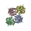
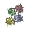
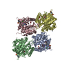
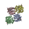
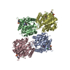
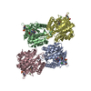
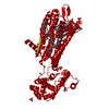
CHAIN A IS AN INTERNAL FUSION OF LYSOZYME (RESIDUES 2-161 OF UNP P00720) BETWEEN RESIDUES 52-263 ...CHAIN A IS AN INTERNAL FUSION OF LYSOZYME (RESIDUES 2-161 OF UNP P00720) BETWEEN RESIDUES 52-263 AND RESIDUES 270-352 OF MU-TYPE OPIOID RECEPTOR (UNP P42866). AN OFFSET OF 1000 HAS BEEN ADDED TO LYSOZYME RESIDUE NUMBERS WITHIN THE COORDINATES TO DISTINGUISH THAT PORTION OF CHAIN A. LYSOZYME RESIDUES ARE THEREFORE NUMBERED 1002-1161.

-
Experimental details

-
Experiment

ExperimentMethod: X-RAY DIFFRACTION / Number of used crystals: 25

-
Sample preparation

CrystalDensity Matthews: 3.82 Å3/Da / Density % sol: 67.78 %
Crystal growTemperature: 293 K / Method: lipidic cubic phase / pH: 7
Details: 100 mM HEPES, pH 7.0, 300 mM lithium sulfate, 7.5% DMSO, 30-38% PEG400 in monoolein:cholesterol mixed in a 10:1 ratio, LIPIDIC CUBIC PHASE, temperature 293K

-
Data collection

DiffractionMean temperature: 78 K
Diffraction sourceSource: SYNCHROTRON / Site: APS / Beamline: 23-ID-D / Wavelength: 1.033 Å
DetectorType: MARMOSAIC 300 mm CCD / Detector: CCD / Date: Nov 30, 2011 / Details: Mirrors
RadiationMonochromator: Double crystal cryo-cooled Si(111) / Protocol: SINGLE WAVELENGTH / Monochromatic (M) / Laue (L): M / Scattering type: x-ray
Radiation wavelengthWavelength: 1.033 Å / Relative weight: 1
ReflectionResolution: 2.8→30.5 Å / Num. obs: 19145 / % possible obs: 99 % / Observed criterion σ(I): -3 / Redundancy: 5.8 % / Rmerge(I) obs: 0.144 / Net I/σ(I): 10.6
Reflection shellResolution: 2.8→2.9 Å / Redundancy: 5.5 % / Rmerge(I) obs: 0.788 / Mean I/σ(I) obs: 1.8 / Num. unique all: 1907 / % possible all: 99.3

-
Processing

Software
NameVersionClassification
Blu-Icedata collection
PHASERphasing
PHENIX(phenix.refine: 1.7.2_869)refinement
HKL-2000data reduction
HKL-2000data scaling
RefinementMethod to determine structure: MOLECULAR REPLACEMENT
Starting model: PDB ENTRY 3ODU
Resolution: 2.8→30.488 Å / SU ML: 0.86 / Cross valid method: THROUGHOUT / σ(F): 1.37 / Phase error: 29.73 / Stereochemistry target values: ML
RfactorNum. reflection% reflectionSelection details
Rfree0.2753 946 4.99 %RANDOM
Rwork0.2326 ---
obs0.2347 18974 97.54 %-
Solvent computationShrinkage radii: 0.9 Å / VDW probe radii: 1.11 Å / Solvent model: FLAT BULK SOLVENT MODEL / Bsol: 59.885 Å2 / ksol: 0.319 e/Å3
Displacement parameters
Baniso -1Baniso -2Baniso -3
1-23.1886 Å20 Å29.0174 Å2
2---18.4026 Å20 Å2
3----4.786 Å2
Refinement stepCycle: LAST / Resolution: 2.8→30.488 Å
ProteinNucleic acidLigandSolventTotal
Num. atoms3476 0 177 37 3690
Refine LS restraints
Refine-IDTypeDev idealNumber
X-RAY DIFFRACTIONf_bond_d0.0053727
X-RAY DIFFRACTIONf_angle_d0.9285083
X-RAY DIFFRACTIONf_dihedral_angle_d12.6151338
X-RAY DIFFRACTIONf_chiral_restr0.057597
X-RAY DIFFRACTIONf_plane_restr0.004599
LS refinement shell
Resolution (Å)Rfactor RfreeNum. reflection RfreeRfactor RworkNum. reflection RworkRefine-ID% reflection obs (%)
2.8-2.94750.36211220.32952355X-RAY DIFFRACTION89
2.9475-3.1320.33181390.28972614X-RAY DIFFRACTION99
3.132-3.37360.32321360.26892603X-RAY DIFFRACTION99
3.3736-3.71260.30521360.23282607X-RAY DIFFRACTION99
3.7126-4.24850.23811390.20892605X-RAY DIFFRACTION99
4.2485-5.3480.24451360.21762608X-RAY DIFFRACTION99
5.348-30.48970.26951380.21572636X-RAY DIFFRACTION98
Refinement TLS params.

Method: refined / Refine-ID: X-RAY DIFFRACTION

IDL112)L122)L132)L222)L232)L332)S11 (Å °)S12 (Å °)S13 (Å °)S21 (Å °)S22 (Å °)S23 (Å °)S31 (Å °)S32 (Å °)S33 (Å °)T112)T122)T132)T222)T232)T332)Origin x (Å)Origin y (Å)Origin z (Å)
11.7115-0.3363-0.42633.69220.65343.3722-0.0406-0.02320.0371-0.12030.056-0.3849-0.06520.13810.00030.092-0.01530.01530.23980.01680.2363-18.86890.3792-13.7282
22.7066-2.8011-1.0583.8359-0.21112.0678-0.06660.0159-0.3047-0.2615-0.0279-0.18640.53010.25860.0020.36790.02210.00690.34660.00470.3573-26.0211-11.7763-4.0518
35.15-3.02031.41125.8439-1.64963.78260.794-1.0357-0.461.508-0.1370.05130.71450.5213-0.57141.2177-0.1736-0.07160.7202-0.00910.6193-17.889128.026618.6757
43.85961.7370.46627.75721.06193.98450.3453-0.0306-0.16110.8023-0.52421.5999-0.2786-0.26810.18690.4854-0.08350.01530.3347-0.11410.6684-23.720546.55477.7946
52.8433-0.2032-1.17832.49430.72535.2252-0.41710.3121-0.0144-1.26450.35060.10420.109-0.1192-0.00030.2911-0.0148-0.0760.36950.02520.2611-31.20671.4132-15.7628
Refinement TLS group
IDRefine-IDRefine TLS-IDSelection detailsAuth asym-IDAuth seq-ID
1X-RAY DIFFRACTION1chain 'A' and (resseq 65:205)A65 - 205
2X-RAY DIFFRACTION2chain 'A' and (resseq 206:258)A206 - 258
3X-RAY DIFFRACTION3chain 'A' and (resseq 259:263 or resseq 1002:1070)A259 - 263
4X-RAY DIFFRACTION3chain 'A' and (resseq 259:263 or resseq 1002:1070)A264 - 1070
5X-RAY DIFFRACTION4chain 'A' and (resseq 1071:1161 or resseq 270:272)A333 - 1161
6X-RAY DIFFRACTION4chain 'A' and (resseq 1071:1161 or resseq 270:272)A270 - 426
7X-RAY DIFFRACTION5chain 'A' and (resseq 273:352)A273 - 506

+
About Yorodumi

-
News

-
Feb 9, 2022. New format data for meta-information of EMDB entries

New format data for meta-information of EMDB entries

  • Version 3 of the EMDB header file is now the official format.
  • The previous official version 1.9 will be removed from the archive.

Related info.:EMDB header

External links:wwPDB to switch to version 3 of the EMDB data model

-
Aug 12, 2020. Covid-19 info

Covid-19 info

URL: https://pdbj.org/emnavi/covid19.php

New page: Covid-19 featured information page in EM Navigator.

Related info.:Covid-19 info / Mar 5, 2020. Novel coronavirus structure data

+
Mar 5, 2020. Novel coronavirus structure data

Novel coronavirus structure data

Related info.:Yorodumi Speices / Aug 12, 2020. Covid-19 info

External links:COVID-19 featured content - PDBj / Molecule of the Month (242):Coronavirus Proteases

+
Jan 31, 2019. EMDB accession codes are about to change! (news from PDBe EMDB page)

EMDB accession codes are about to change! (news from PDBe EMDB page)

  • The allocation of 4 digits for EMDB accession codes will soon come to an end. Whilst these codes will remain in use, new EMDB accession codes will include an additional digit and will expand incrementally as the available range of codes is exhausted. The current 4-digit format prefixed with “EMD-” (i.e. EMD-XXXX) will advance to a 5-digit format (i.e. EMD-XXXXX), and so on. It is currently estimated that the 4-digit codes will be depleted around Spring 2019, at which point the 5-digit format will come into force.
  • The EM Navigator/Yorodumi systems omit the EMD- prefix.

Related info.:Q: What is EMD? / ID/Accession-code notation in Yorodumi/EM Navigator

External links:EMDB Accession Codes are Changing Soon! / Contact to PDBj

+
Jul 12, 2017. Major update of PDB

Major update of PDB

  • wwPDB released updated PDB data conforming to the new PDBx/mmCIF dictionary.
  • This is a major update changing the version number from 4 to 5, and with Remediation, in which all the entries are updated.
  • In this update, many items about electron microscopy experimental information are reorganized (e.g. em_software).
  • Now, EM Navigator and Yorodumi are based on the updated data.

External links:wwPDB Remediation / Enriched Model Files Conforming to OneDep Data Standards Now Available in the PDB FTP Archive

-
Yorodumi

Thousand views of thousand structures

  • Yorodumi is a browser for structure data from EMDB, PDB, SASBDB, etc.
  • This page is also the successor to EM Navigator detail page, and also detail information page/front-end page for Omokage search.
  • The word "yorodu" (or yorozu) is an old Japanese word meaning "ten thousand". "mi" (miru) is to see.

Related info.:EMDB / PDB / SASBDB / Comparison of 3 databanks / Yorodumi Search / Aug 31, 2016. New EM Navigator & Yorodumi / Yorodumi Papers / Jmol/JSmol / Function and homology information / Changes in new EM Navigator and Yorodumi

Read more