 Movie
Movie Controller


+ Open data

- Basic information
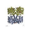
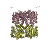
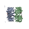
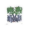
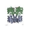
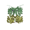
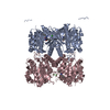
| Entry | Database: PDB / ID: 5fuh | ||||||
|---|---|---|---|---|---|---|---|
| Title | Pseudomonas aeruginosa RmlA in complex with allosteric inhibitor | ||||||
 Components Components | GLUCOSE-1-PHOSPHATE THYMIDYLYLTRANSFERASE | ||||||
 Keywords Keywords | TRANSFERASE / THYMIDYLYL / ALLOSTERIC / INHIBITOR / PSEUDOMONAS | ||||||
| Function / homology |  Function and homology information Function and homology informationglucose-1-phosphate thymidylyltransferase / glucose-1-phosphate thymidylyltransferase activity / dTDP-rhamnose biosynthetic process / lipopolysaccharide core region biosynthetic process / polysaccharide biosynthetic process / nucleotide binding / metal ion binding / cytosol Similarity search - Function | ||||||
| Biological species |   PSEUDOMONAS AERUGINOSA (bacteria) PSEUDOMONAS AERUGINOSA (bacteria) | ||||||
| Method |  X-RAY DIFFRACTION / SYNCHROTRON / MOLECULAR REPLACEMENT / Resolution: 1.6 Å X-RAY DIFFRACTION / SYNCHROTRON / MOLECULAR REPLACEMENT / Resolution: 1.6 Å | ||||||
 Authors Authors | Alphey, M.S. / Tran, F. / Westwood, N.J. / Naismith, J.H. | ||||||
 Citation Citation | Journal: To be Published Title: Allosteric Competitive Inhibitors of the Glucose-1-Phosphate Thymidylyltransferase (Rmla) from Pseudomonas Aeruginosa. Authors: Tran, F. / Alphey, M.S. / Westwood, N.J. / Naismith, J.H. | ||||||
| History |
|
- Structure visualization
| Structure viewer | Molecule: MolmilJmol/JSmol |
|---|
- Downloads & links
-Download
| PDBx/mmCIF format | 5fuh.cif.gz | 254.3 KB | Display | PDBx/mmCIF format |
|---|---|---|---|---|
| PDB format | pdb5fuh.ent.gz | 207 KB | Display | PDB format |
| PDBx/mmJSON format | 5fuh.json.gz | Tree view | PDBx/mmJSON format | |
| Others |  Other downloads Other downloads |
-Validation report
| Arichive directory | https://data.pdbj.org/pub/pdb/validation_reports/fu/5fuhftp://data.pdbj.org/pub/pdb/validation_reports/fu/5fuh | HTTPS FTP |
|---|
-Related structure data
| Related structure data |  5ftsC  5ftvC 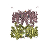 5fu0C 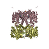 5fu8C 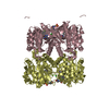 5fyeC 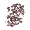 4asjS C: citing same article ( S: Starting model for refinement |
|---|---|
| Similar structure data |


-Links
 PDBj
PDBj

- Assembly
| Deposited unit | 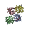
| ||||||||||||||||||||||||
|---|---|---|---|---|---|---|---|---|---|---|---|---|---|---|---|---|---|---|---|---|---|---|---|---|---|
| 1 | 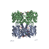
| ||||||||||||||||||||||||
| 2 | 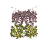
| ||||||||||||||||||||||||
| Unit cell |
| ||||||||||||||||||||||||
| Components on special symmetry positions |
| ||||||||||||||||||||||||
| Noncrystallographic symmetry (NCS) | NCS oper:
|
-Components
-Protein , 1 types, 4 molecules ABCD
| #1: Protein | Mass: 33664.121 Da / Num. of mol.: 4 Source method: isolated from a genetically manipulated source Source: (gene. exp.) PSEUDOMONAS AERUGINOSA (bacteria) / Production host: References: UniProt: Q9HU22, glucose-1-phosphate thymidylyltransferase |
|---|
-Non-polymers , 5 types, 588 molecules 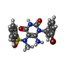


| #2: Chemical | ChemComp-HKX /  Mass: 465.321 Da / Num. of mol.: 4 / Source method: obtained synthetically / Formula: C18H17BrN4O4S Mass: 465.321 Da / Num. of mol.: 4 / Source method: obtained synthetically / Formula: C18H17BrN4O4S#3: Chemical | ChemComp-MES /  Mass: 195.237 Da / Num. of mol.: 4 / Source method: obtained synthetically / Formula: C6H13NO4S / Comment: pH buffer*YM Mass: 195.237 Da / Num. of mol.: 4 / Source method: obtained synthetically / Formula: C6H13NO4S / Comment: pH buffer*YM#4: Chemical | ChemComp-CL /  Mass: 35.453 Da / Num. of mol.: 4 / Source method: obtained synthetically / Formula: Cl Mass: 35.453 Da / Num. of mol.: 4 / Source method: obtained synthetically / Formula: Cl#5: Chemical | ChemComp-GOL /  Mass: 92.094 Da / Num. of mol.: 4 / Source method: obtained synthetically / Formula: C3H8O3 Mass: 92.094 Da / Num. of mol.: 4 / Source method: obtained synthetically / Formula: C3H8O3#6: Water | ChemComp-HOH / | Mass: 18.015 Da / Num. of mol.: 572 / Source method: isolated from a natural source / Formula: H2O |
|---|
-Details
| Sequence details | N-TERMINAL HIS-TAG PRESENT |
|---|
-Experimental details
-Experiment
| Experiment | Method: X-RAY DIFFRACTION / Number of used crystals: 1 |
|---|
- Sample preparation
| Crystal | Density Matthews: 2.59 Å3/Da / Density % sol: 52.58 % / Description: NONE |
|---|---|
| Crystal grow | pH: 6 Details: 4% PEG 6000, 0.1 M MES PH 6, 0.05 M MGCL2, 0.1 M NA BR, 1% BETA-MERCAPTOETHANOL |
-Data collection
| Diffraction | Mean temperature: 100 K |
|---|---|
| Diffraction source | Source: SYNCHROTRON / Site: Diamond  / Beamline: I04 / Wavelength: 0.9795 / Beamline: I04 / Wavelength: 0.9795 |
| Detector | Type: DECTRIS PILATUS 6M / Detector: PIXEL / Date: Dec 10, 2014 / Details: MIRRORS |
| Radiation | Monochromator: DOUBLE CRYSTAL / Protocol: SINGLE WAVELENGTH / Monochromatic (M) / Laue (L): M / Scattering type: x-ray |
| Radiation wavelength | Wavelength: 0.9795 Å / Relative weight: 1 |
| Reflection | Resolution: 1.59→67.35 Å / Num. obs: 175397 / % possible obs: 99.1 % / Observed criterion σ(I): 2 / Redundancy: 6 % / Biso Wilson estimate: 22.5 Å2 / Rmerge(I) obs: 0.1 / Net I/σ(I): 10.5 |
| Reflection shell | Resolution: 1.59→1.63 Å / Redundancy: 3.5 % / Rmerge(I) obs: 0.68 / Mean I/σ(I) obs: 2.1 / % possible all: 96.1 |
- Processing
| Software |
| ||||||||||||||||||||||||||||||||||||||||||||||||||||||||||||||||||||||||||||||||||||||||||||||||||||||||||||||||||||||||||||||||||||||||||||||||||||||||||||||||||||||||||||||||||||||
|---|---|---|---|---|---|---|---|---|---|---|---|---|---|---|---|---|---|---|---|---|---|---|---|---|---|---|---|---|---|---|---|---|---|---|---|---|---|---|---|---|---|---|---|---|---|---|---|---|---|---|---|---|---|---|---|---|---|---|---|---|---|---|---|---|---|---|---|---|---|---|---|---|---|---|---|---|---|---|---|---|---|---|---|---|---|---|---|---|---|---|---|---|---|---|---|---|---|---|---|---|---|---|---|---|---|---|---|---|---|---|---|---|---|---|---|---|---|---|---|---|---|---|---|---|---|---|---|---|---|---|---|---|---|---|---|---|---|---|---|---|---|---|---|---|---|---|---|---|---|---|---|---|---|---|---|---|---|---|---|---|---|---|---|---|---|---|---|---|---|---|---|---|---|---|---|---|---|---|---|---|---|---|---|
| Refinement | Method to determine structure: MOLECULAR REPLACEMENT Starting model: PDB ENTRY 4ASJ Resolution: 1.6→67.349 Å / Cor.coef. Fo:Fc: 0.959 / Cor.coef. Fo:Fc free: 0.95 / SU B: 1.997 / SU ML: 0.07 / Cross valid method: THROUGHOUT / ESU R: 0.094 / ESU R Free: 0.092 / Stereochemistry target values: MAXIMUM LIKELIHOOD Details: HYDROGENS HAVE BEEN ADDED IN THE RIDING POSITIONS. SOME RESIDUES AT THE N-TERMINUS ARE DISORDERED. SOME RESIDUES HAVE BEEN MODELLED IN MULTIPLE CONFORMATIONS. THE BOUND INHIBITOR HAS PARTIAL ...Details: HYDROGENS HAVE BEEN ADDED IN THE RIDING POSITIONS. SOME RESIDUES AT THE N-TERMINUS ARE DISORDERED. SOME RESIDUES HAVE BEEN MODELLED IN MULTIPLE CONFORMATIONS. THE BOUND INHIBITOR HAS PARTIAL OCCUPANCY. SOME EXTRA ELECTRON DENSITY IS OBSERVED IN THE PARTIALLY OCCUPIED INHIBITOR BINDING SITE, MOST LIKELY DUE TO WATERS BOUND WHEN INHIBITOR IS ABSENT. SOME EXTRA ELECTRON DENSITY IS OBSERVED NEAR THE MULTIPLE CONFORMATIONS OF GLN237, MOST LIKELY DUE TO WATERS BOUND WHEN SIDE CHAIN IS IN ALTERNATIVE CONFORMATION.
| ||||||||||||||||||||||||||||||||||||||||||||||||||||||||||||||||||||||||||||||||||||||||||||||||||||||||||||||||||||||||||||||||||||||||||||||||||||||||||||||||||||||||||||||||||||||
| Solvent computation | Ion probe radii: 0.4 Å / VDW probe radii: 1.2 Å / Solvent model: BABINET MODEL PLUS MASK | ||||||||||||||||||||||||||||||||||||||||||||||||||||||||||||||||||||||||||||||||||||||||||||||||||||||||||||||||||||||||||||||||||||||||||||||||||||||||||||||||||||||||||||||||||||||
| Displacement parameters | Biso mean: 29.619 Å2
| ||||||||||||||||||||||||||||||||||||||||||||||||||||||||||||||||||||||||||||||||||||||||||||||||||||||||||||||||||||||||||||||||||||||||||||||||||||||||||||||||||||||||||||||||||||||
| Refinement step | Cycle: LAST / Resolution: 1.6→67.349 Å
| ||||||||||||||||||||||||||||||||||||||||||||||||||||||||||||||||||||||||||||||||||||||||||||||||||||||||||||||||||||||||||||||||||||||||||||||||||||||||||||||||||||||||||||||||||||||
| Refine LS restraints |
|