 Movie
Movie Controller


+ Open data

- Basic information
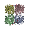
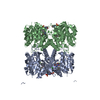
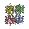
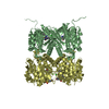
| Entry | Database: PDB / ID: 5fu0 | ||||||
|---|---|---|---|---|---|---|---|
| Title | Pseudomonas aeruginosa RmlA in complex with allosteric inhibitor | ||||||
 Components Components | GLUCOSE-1-PHOSPHATE THYMIDYLYLTRANSFERASE | ||||||
 Keywords Keywords | TRANSFERASE / THYMIDYLYL / ALLOSTERIC / INHIBITOR / PSEUDOMONAS | ||||||
| Function / homology |  Function and homology information Function and homology informationglucose-1-phosphate thymidylyltransferase / glucose-1-phosphate thymidylyltransferase activity / dTDP-rhamnose biosynthetic process / lipopolysaccharide core region biosynthetic process / polysaccharide biosynthetic process / nucleotide binding / metal ion binding / cytosol Similarity search - Function | ||||||
| Biological species |   PSEUDOMONAS AERUGINOSA (bacteria) PSEUDOMONAS AERUGINOSA (bacteria) | ||||||
| Method |  X-RAY DIFFRACTION / MOLECULAR REPLACEMENT / Resolution: 1.9 Å X-RAY DIFFRACTION / MOLECULAR REPLACEMENT / Resolution: 1.9 Å | ||||||
 Authors Authors | Alphey, M.S. / Tran, F. / Westwood, N.J. / Naismith, J.H. | ||||||
 Citation Citation | Journal: To be Published Title: Allosteric Competitive Inhibitors of the Glucose-1-Phosphate Thymidylyltransferase (Rmla) from Pseudomonas Aeruginosa. Authors: Tran, F. / Alphey, M.S. / Westwood, N.J. / Naismith, J.H. | ||||||
| History |
|
- Structure visualization
| Structure viewer | Molecule: MolmilJmol/JSmol |
|---|
- Downloads & links
-Download
| PDBx/mmCIF format | 5fu0.cif.gz | 260.9 KB | Display | PDBx/mmCIF format |
|---|---|---|---|---|
| PDB format | pdb5fu0.ent.gz | 212 KB | Display | PDB format |
| PDBx/mmJSON format | 5fu0.json.gz | Tree view | PDBx/mmJSON format | |
| Others |  Other downloads Other downloads |
-Validation report
| Arichive directory | https://data.pdbj.org/pub/pdb/validation_reports/fu/5fu0ftp://data.pdbj.org/pub/pdb/validation_reports/fu/5fu0 | HTTPS FTP |
|---|
-Related structure data
| Related structure data |  5ftsC 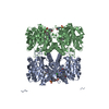 5ftvC 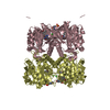 5fu8C 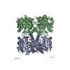 5fuhC 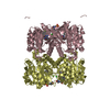 5fyeC 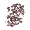 4asjS S: Starting model for refinement C: citing same article ( |
|---|---|
| Similar structure data |
-Links
 PDBj
PDBj

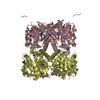
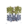
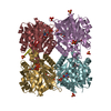
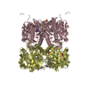
- Assembly
| Deposited unit | 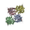
| ||||||||||||||||
|---|---|---|---|---|---|---|---|---|---|---|---|---|---|---|---|---|---|
| 1 | 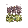
| ||||||||||||||||
| 2 | 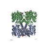
| ||||||||||||||||
| Unit cell |
| ||||||||||||||||
| Noncrystallographic symmetry (NCS) | NCS oper:
|
-Components
| #1: Protein | Mass: 33664.121 Da / Num. of mol.: 4 Source method: isolated from a genetically manipulated source Source: (gene. exp.) PSEUDOMONAS AERUGINOSA (bacteria) / Production host: References: UniProt: Q9HU22, glucose-1-phosphate thymidylyltransferase #2: Chemical | ChemComp-FKH / 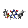  Mass: 400.452 Da / Num. of mol.: 4 / Source method: obtained synthetically / Formula: C19H20N4O4S Mass: 400.452 Da / Num. of mol.: 4 / Source method: obtained synthetically / Formula: C19H20N4O4S#3: Chemical | ChemComp-MES /   Mass: 195.237 Da / Num. of mol.: 4 / Source method: obtained synthetically / Formula: C6H13NO4S / Comment: pH buffer*YM Mass: 195.237 Da / Num. of mol.: 4 / Source method: obtained synthetically / Formula: C6H13NO4S / Comment: pH buffer*YM#4: Chemical | ChemComp-CL /   Mass: 35.453 Da / Num. of mol.: 4 / Source method: obtained synthetically / Formula: Cl Mass: 35.453 Da / Num. of mol.: 4 / Source method: obtained synthetically / Formula: Cl#5: Water | ChemComp-HOH / |  Mass: 18.015 Da / Num. of mol.: 974 / Source method: isolated from a natural source / Formula: H2O Mass: 18.015 Da / Num. of mol.: 974 / Source method: isolated from a natural source / Formula: H2OSequence details | N-TERMINAL HIS-TAG PRESENT | |
|---|
-Experimental details
-Experiment
| Experiment | Method: X-RAY DIFFRACTION / Number of used crystals: 1 |
|---|
- Sample preparation
| Crystal | Density Matthews: 2.56 Å3/Da / Density % sol: 52.07 % / Description: NONE |
|---|---|
| Crystal grow | pH: 6 Details: 4% PEG 6000, 0.1 M MES PH 6, 0.05 M MGCL2, 0.1 M NA BR, 1% BETA-MERCAPTOETHANOL |
-Data collection
| Diffraction | Mean temperature: 100 K |
|---|---|
| Diffraction source | Source: ROTATING ANODE / Type: RIGAKU MICROMAX-007 HF / Wavelength: 1.5418 |
| Detector | Type: MSC SATURN 944 / Detector: CCD / Date: Dec 21, 2015 / Details: MIRRORS |
| Radiation | Protocol: SINGLE WAVELENGTH / Monochromatic (M) / Laue (L): M / Scattering type: x-ray |
| Radiation wavelength | Wavelength: 1.5418 Å / Relative weight: 1 |
| Reflection | Resolution: 1.9→27.7 Å / Num. obs: 102239 / % possible obs: 93 % / Observed criterion σ(I): 2 / Redundancy: 3.8 % / Biso Wilson estimate: 16.17 Å2 / Rmerge(I) obs: 0.1 / Net I/σ(I): 7.1 |
| Reflection shell | Resolution: 1.9→2 Å / Redundancy: 2.5 % / Rmerge(I) obs: 0.32 / Mean I/σ(I) obs: 3.3 / % possible all: 79 |
- Processing
| Software |
| ||||||||||||||||||||||||||||||||||||||||||||||||||||||||||||||||||||||||||||||||||||||||||||||||||||||||||||||||||||||||||||||||||||||||||||||||||||||||||||||||||||||||||||||||||||||
|---|---|---|---|---|---|---|---|---|---|---|---|---|---|---|---|---|---|---|---|---|---|---|---|---|---|---|---|---|---|---|---|---|---|---|---|---|---|---|---|---|---|---|---|---|---|---|---|---|---|---|---|---|---|---|---|---|---|---|---|---|---|---|---|---|---|---|---|---|---|---|---|---|---|---|---|---|---|---|---|---|---|---|---|---|---|---|---|---|---|---|---|---|---|---|---|---|---|---|---|---|---|---|---|---|---|---|---|---|---|---|---|---|---|---|---|---|---|---|---|---|---|---|---|---|---|---|---|---|---|---|---|---|---|---|---|---|---|---|---|---|---|---|---|---|---|---|---|---|---|---|---|---|---|---|---|---|---|---|---|---|---|---|---|---|---|---|---|---|---|---|---|---|---|---|---|---|---|---|---|---|---|---|---|
| Refinement | Method to determine structure: MOLECULAR REPLACEMENT Starting model: PDB ENTRY 4ASJ Resolution: 1.9→134.681 Å / Cor.coef. Fo:Fc: 0.881 / Cor.coef. Fo:Fc free: 0.855 / SU B: 4.438 / SU ML: 0.125 / Cross valid method: THROUGHOUT / ESU R: 0.23 / ESU R Free: 0.191 / Stereochemistry target values: MAXIMUM LIKELIHOOD Details: HYDROGENS HAVE BEEN ADDED IN THE RIDING POSITIONS. SOME RESIDUES AT N-TERMINI OF SUBUNITS ARE DISORDERED
| ||||||||||||||||||||||||||||||||||||||||||||||||||||||||||||||||||||||||||||||||||||||||||||||||||||||||||||||||||||||||||||||||||||||||||||||||||||||||||||||||||||||||||||||||||||||
| Solvent computation | Ion probe radii: 0.4 Å / VDW probe radii: 1.2 Å / Solvent model: BABINET MODEL PLUS MASK | ||||||||||||||||||||||||||||||||||||||||||||||||||||||||||||||||||||||||||||||||||||||||||||||||||||||||||||||||||||||||||||||||||||||||||||||||||||||||||||||||||||||||||||||||||||||
| Displacement parameters | Biso mean: 25.991 Å2
| ||||||||||||||||||||||||||||||||||||||||||||||||||||||||||||||||||||||||||||||||||||||||||||||||||||||||||||||||||||||||||||||||||||||||||||||||||||||||||||||||||||||||||||||||||||||
| Refinement step | Cycle: LAST / Resolution: 1.9→134.681 Å
| ||||||||||||||||||||||||||||||||||||||||||||||||||||||||||||||||||||||||||||||||||||||||||||||||||||||||||||||||||||||||||||||||||||||||||||||||||||||||||||||||||||||||||||||||||||||
| Refine LS restraints |
|