 Movie
Movie Controller

[English] 日本語
 Yorodumi
- PDB-8qmb: Nucleant-assisted 2.0 A resolution structure of the Streptococcus... -
+ Open data

- Basic information
| Entry | Database: PDB / ID: 8qmb | ||||||
|---|---|---|---|---|---|---|---|
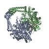
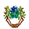
| Title | Nucleant-assisted 2.0 A resolution structure of the Streptococcus pneumoniae topoisomerase IV-V18mer DNA complex with the novel fluoroquinolone Delafloxacin | ||||||
 Components Components |
| ||||||
 Keywords Keywords | ISOMERASE / PROTEIN-DNA CLEAVAGE COMPLEX / TOPOISOMERASE IIA / DELAFLOXACIN / TOPOISOMERASE IV-DNA-ANTIBIOTIC COMPLEX / PEGylated GRAPHENE | ||||||
| Function / homology |  Function and homology information Function and homology informationDNA topoisomerase type II (double strand cut, ATP-hydrolyzing) activity / DNA topoisomerase (ATP-hydrolysing) / extrinsic component of plasma membrane / DNA topological change / chromosome segregation / chromosome / DNA binding / ATP binding / metal ion binding Similarity search - Function | ||||||
| Biological species |   Streptococcus pneumoniae (bacteria) Streptococcus pneumoniae (bacteria) | ||||||
| Method |  X-RAY DIFFRACTION / SYNCHROTRON / MOLECULAR REPLACEMENT / Resolution: 2 Å X-RAY DIFFRACTION / SYNCHROTRON / MOLECULAR REPLACEMENT / Resolution: 2 Å | ||||||
 Authors Authors | Najmudin, S. / Pan, X.S. / Wang, B. / Chayen, N.E. / Fisher, L.M. / Sanderson, M.R. | ||||||
| Funding support |  United Kingdom, 1items United Kingdom, 1items
| ||||||
 Citation Citation | Journal: To Be Published Title: The nature of the molecular interactions at high resolution of the Streptococcus pneumoniae topoisomerase IV-DNA complex with the novel fluoroquinolone Delafloxacin. Authors: Najmudin, S. / Pan, X.S. / Wang, B. / Chayen, N.E. / Fisher, L.M. / Sanderson, M.R. | ||||||
| History |
|
- Structure visualization
| Structure viewer | Molecule: MolmilJmol/JSmol |
|---|
- Downloads & links
-Download
| PDBx/mmCIF format | 8qmb.cif.gz | 1.4 MB | Display | PDBx/mmCIF format |
|---|---|---|---|---|
| PDB format | pdb8qmb.ent.gz | 1001.1 KB | Display | PDB format |
| PDBx/mmJSON format | 8qmb.json.gz | Tree view | PDBx/mmJSON format | |
| Others |  Other downloads Other downloads |
-Validation report
| Summary document | 8qmb_validation.pdf.gz | 1.3 MB | Display | wwPDB validaton report |
|---|---|---|---|---|
| Full document | 8qmb_full_validation.pdf.gz | 1.4 MB | Display | |
| Data in XML | 8qmb_validation.xml.gz | 77.7 KB | Display | |
| Data in CIF | 8qmb_validation.cif.gz | 106.2 KB | Display | |
| Arichive directory | https://data.pdbj.org/pub/pdb/validation_reports/qm/8qmbftp://data.pdbj.org/pub/pdb/validation_reports/qm/8qmb | HTTPS FTP |
-Related structure data
-Links
 PDBj
PDBj


- Assembly
| Deposited unit | 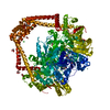
| ||||||||||||
|---|---|---|---|---|---|---|---|---|---|---|---|---|---|
| 1 |
| ||||||||||||
| Unit cell |
| ||||||||||||
| Noncrystallographic symmetry (NCS) | NCS domain:
NCS domain segments: Component-ID: 1 / Ens-ID: 1 / Beg auth comp-ID: LEU / Beg label comp-ID: LEU / End auth comp-ID: ALA / End label comp-ID: ALA / Auth asym-ID: A / Label asym-ID: A / Auth seq-ID: 411 - 1486 / Label seq-ID: 9 - 732
NCS ensembles : (Details: Local NCS retraints between domains: 1 2) |
-Components
-Protein , 1 types, 2 molecules AB
| #1: Protein | Mass: 84211.422 Da / Num. of mol.: 2 / Mutation: Insertion of His at postion 648 Source method: isolated from a genetically manipulated source Details: The fused TOPOISOMERASE IV CLEAVAGE COMPLEX comprises the C-terminal domain of the ParE30 domain (residues 415-647), a His insert at position 648 and the N-terminal domain of ParC55 ...Details: The fused TOPOISOMERASE IV CLEAVAGE COMPLEX comprises the C-terminal domain of the ParE30 domain (residues 415-647), a His insert at position 648 and the N-terminal domain of ParC55 (residues 1001-1486),The fused TOPOISOMERASE IV CLEAVAGE COMPLEX comprises the C-terminal domain of the ParE30 domain (residues 415-647), a His insert at position 648 and the N-terminal domain of ParC55 (residues 1001-1486),The fused TOPOISOMERASE IV CLEAVAGE COMPLEX comprises the C-terminal domain of the ParE30 domain (residues 415-647), a His insert at position 648 and the N-terminal domain of ParC55 (residues 1001-1486),The fused TOPOISOMERASE IV CLEAVAGE COMPLEX comprises the C-terminal domain of the ParE30 domain (residues 415-647), a His insert at position 648 and the N-terminal domain of ParC55 (residues 1001-1486) Source: (gene. exp.) Streptococcus pneumoniae (bacteria) / Strain: 7785 / Gene: parE, SP_0852, parC, SP_0855 / Plasmid: PET29A / Production host: References: UniProt: Q59961, UniProt: P72525, DNA topoisomerase (ATP-hydrolysing) |
|---|
-DNA chain , 4 types, 4 molecules EFGH
| #2: DNA chain | Mass: 2168.445 Da / Num. of mol.: 1 / Source method: obtained synthetically / Details: plamid pBR322 / Source: (synth.) |
|---|---|
| #3: DNA chain | Mass: 3333.198 Da / Num. of mol.: 1 / Source method: obtained synthetically / Details: plasmid pBR322 / Source: (synth.) |
| #4: DNA chain | Mass: 2121.436 Da / Num. of mol.: 1 / Source method: obtained synthetically / Details: plasmid pBR322 / Source: (synth.) |
| #5: DNA chain | Mass: 3317.199 Da / Num. of mol.: 1 / Source method: obtained synthetically / Details: plasmid pBR322 / Source: (synth.) |
-Non-polymers , 8 types, 1082 molecules 


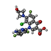

| #6: Chemical | ChemComp-MG /  Mass: 24.305 Da / Num. of mol.: 6 / Source method: obtained synthetically / Formula: Mg Mass: 24.305 Da / Num. of mol.: 6 / Source method: obtained synthetically / Formula: Mg#7: Chemical |  Mass: 35.453 Da / Num. of mol.: 2 / Source method: obtained synthetically / Formula: Cl Mass: 35.453 Da / Num. of mol.: 2 / Source method: obtained synthetically / Formula: Cl#8: Chemical |  Mass: 118.174 Da / Num. of mol.: 3 / Source method: obtained synthetically / Formula: C6H14O2 / Comment: precipitant*YM Mass: 118.174 Da / Num. of mol.: 3 / Source method: obtained synthetically / Formula: C6H14O2 / Comment: precipitant*YM#9: Chemical |  Mass: 104.061 Da / Num. of mol.: 2 / Source method: obtained synthetically / Formula: C3H4O4 Mass: 104.061 Da / Num. of mol.: 2 / Source method: obtained synthetically / Formula: C3H4O4#10: Chemical | ChemComp-ACT /  Mass: 59.044 Da / Num. of mol.: 4 / Source method: obtained synthetically / Formula: C2H3O2 Mass: 59.044 Da / Num. of mol.: 4 / Source method: obtained synthetically / Formula: C2H3O2#11: Chemical | ChemComp-FMT /  Mass: 46.025 Da / Num. of mol.: 4 / Source method: obtained synthetically / Formula: CH2O2 Mass: 46.025 Da / Num. of mol.: 4 / Source method: obtained synthetically / Formula: CH2O2#12: Chemical |  Mass: 440.760 Da / Num. of mol.: 2 / Source method: obtained synthetically / Formula: C18H12ClF3N4O4 / Feature type: SUBJECT OF INVESTIGATION Mass: 440.760 Da / Num. of mol.: 2 / Source method: obtained synthetically / Formula: C18H12ClF3N4O4 / Feature type: SUBJECT OF INVESTIGATION#13: Water | ChemComp-HOH / | Mass: 18.015 Da / Num. of mol.: 1059 / Source method: isolated from a natural source / Formula: H2O |
|---|
-Details
| Has ligand of interest | Y |
|---|
-Experimental details
-Experiment
| Experiment | Method: X-RAY DIFFRACTION / Number of used crystals: 1 |
|---|
- Sample preparation
| Crystal | Density Matthews: 4.22 Å3/Da / Density % sol: 71.1 % |
|---|---|
| Crystal grow | Temperature: 301 K / Method: vapor diffusion, sitting drop / pH: 7 Details: 2.5% Tacsimate, 50 mM Na Cacodylate, 62.5 mM KCl, 7.5 mM MgCl2, 5.5-7.0% Isopropanol, 0.05 mg/mL PEGylated graphene as a nucleant. 30% MPD as cryoprotectant and 1 mM beta-mercaptoethanol in the mother liquor. PH range: 6.5-8.0 |
-Data collection
| Diffraction | Mean temperature: 100 K / Serial crystal experiment: N |
|---|---|
| Diffraction source | Source: SYNCHROTRON / Site: Diamond / Beamline: I04 / Wavelength: 0.9537 Å |
| Detector | Type: DECTRIS EIGER2 XE 16M / Detector: PIXEL / Date: Mar 9, 2023 |
| Radiation | Protocol: SINGLE WAVELENGTH / Monochromatic (M) / Laue (L): M / Scattering type: x-ray |
| Radiation wavelength | Wavelength: 0.9537 Å / Relative weight: 1 |
| Reflection | Resolution: 1.997→83.768 Å / Num. obs: 115609 / % possible obs: 96.6 % / Observed criterion σ(I): 2 / Redundancy: 100.1 % / CC1/2: 1 / Rmerge(I) obs: 0.239 / Rpim(I) all: 0.024 / Rrim(I) all: 0.24 / Net I/σ(I): 19 |
| Reflection shell | Resolution: 1.997→2.306 Å / Redundancy: 82.3 % / Rmerge(I) obs: 2.948 / Mean I/σ(I) obs: 2.5 / Num. unique obs: 6766 / CC1/2: 0.827 / Rpim(I) all: 0.326 / Rrim(I) all: 2.966 / % possible all: 82 |
- Processing
| Software |
| |||||||||||||||||||||||||||||||||||||||||||||||||||||||||||||||||||||||||||||||||||||||||||||||||||||||||||||||||||||||||||||||||||||||||||||||||||||||||||||||||||||||||||||||||||||||||||||||||||||||||||||||||||||||||||||||||||||||
|---|---|---|---|---|---|---|---|---|---|---|---|---|---|---|---|---|---|---|---|---|---|---|---|---|---|---|---|---|---|---|---|---|---|---|---|---|---|---|---|---|---|---|---|---|---|---|---|---|---|---|---|---|---|---|---|---|---|---|---|---|---|---|---|---|---|---|---|---|---|---|---|---|---|---|---|---|---|---|---|---|---|---|---|---|---|---|---|---|---|---|---|---|---|---|---|---|---|---|---|---|---|---|---|---|---|---|---|---|---|---|---|---|---|---|---|---|---|---|---|---|---|---|---|---|---|---|---|---|---|---|---|---|---|---|---|---|---|---|---|---|---|---|---|---|---|---|---|---|---|---|---|---|---|---|---|---|---|---|---|---|---|---|---|---|---|---|---|---|---|---|---|---|---|---|---|---|---|---|---|---|---|---|---|---|---|---|---|---|---|---|---|---|---|---|---|---|---|---|---|---|---|---|---|---|---|---|---|---|---|---|---|---|---|---|---|---|---|---|---|---|---|---|---|---|---|---|---|---|---|---|---|---|
| Refinement | Method to determine structure: MOLECULAR REPLACEMENT / Resolution: 2→83.628 Å / Cor.coef. Fo:Fc: 0.95 / Cor.coef. Fo:Fc free: 0.944 / SU B: 7.887 / SU ML: 0.104 / Cross valid method: FREE R-VALUE / ESU R: 0.204 / ESU R Free: 0.178 Details: Hydrogens have been added in their riding positions
| |||||||||||||||||||||||||||||||||||||||||||||||||||||||||||||||||||||||||||||||||||||||||||||||||||||||||||||||||||||||||||||||||||||||||||||||||||||||||||||||||||||||||||||||||||||||||||||||||||||||||||||||||||||||||||||||||||||||
| Solvent computation | Ion probe radii: 0.8 Å / Shrinkage radii: 0.8 Å / VDW probe radii: 1.2 Å / Solvent model: MASK BULK SOLVENT | |||||||||||||||||||||||||||||||||||||||||||||||||||||||||||||||||||||||||||||||||||||||||||||||||||||||||||||||||||||||||||||||||||||||||||||||||||||||||||||||||||||||||||||||||||||||||||||||||||||||||||||||||||||||||||||||||||||||
| Displacement parameters | Biso mean: 47.121 Å2
| |||||||||||||||||||||||||||||||||||||||||||||||||||||||||||||||||||||||||||||||||||||||||||||||||||||||||||||||||||||||||||||||||||||||||||||||||||||||||||||||||||||||||||||||||||||||||||||||||||||||||||||||||||||||||||||||||||||||
| Refinement step | Cycle: LAST / Resolution: 2→83.628 Å
| |||||||||||||||||||||||||||||||||||||||||||||||||||||||||||||||||||||||||||||||||||||||||||||||||||||||||||||||||||||||||||||||||||||||||||||||||||||||||||||||||||||||||||||||||||||||||||||||||||||||||||||||||||||||||||||||||||||||
| Refine LS restraints |
| |||||||||||||||||||||||||||||||||||||||||||||||||||||||||||||||||||||||||||||||||||||||||||||||||||||||||||||||||||||||||||||||||||||||||||||||||||||||||||||||||||||||||||||||||||||||||||||||||||||||||||||||||||||||||||||||||||||||
| Refine LS restraints NCS |
| |||||||||||||||||||||||||||||||||||||||||||||||||||||||||||||||||||||||||||||||||||||||||||||||||||||||||||||||||||||||||||||||||||||||||||||||||||||||||||||||||||||||||||||||||||||||||||||||||||||||||||||||||||||||||||||||||||||||
| LS refinement shell | Refine-ID: X-RAY DIFFRACTION / Total num. of bins used: 20
| |||||||||||||||||||||||||||||||||||||||||||||||||||||||||||||||||||||||||||||||||||||||||||||||||||||||||||||||||||||||||||||||||||||||||||||||||||||||||||||||||||||||||||||||||||||||||||||||||||||||||||||||||||||||||||||||||||||||
| Refinement TLS params. | Method: refined / Refine-ID: X-RAY DIFFRACTION
| |||||||||||||||||||||||||||||||||||||||||||||||||||||||||||||||||||||||||||||||||||||||||||||||||||||||||||||||||||||||||||||||||||||||||||||||||||||||||||||||||||||||||||||||||||||||||||||||||||||||||||||||||||||||||||||||||||||||
| Refinement TLS group | Selection: ALL |
