[English] 日本語
 Yorodumi
- PDB-8qkk: Cryo-EM structure of MmpL3 from Mycobacterium smegmatis reconstit... -
+ Open data
- Basic information
| Entry | Database: PDB / ID: 8qkk | ||||||||||||
|---|---|---|---|---|---|---|---|---|---|---|---|---|---|
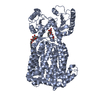
| Title | Cryo-EM structure of MmpL3 from Mycobacterium smegmatis reconstituted into peptidiscs | ||||||||||||
 Components Components | Trehalose monomycolate exporter MmpL3 | ||||||||||||
 Keywords Keywords | MEMBRANE PROTEIN / mycobacterium / trehalose monomycolate / TMM / peptidisc / MmpL3 | ||||||||||||
| Function / homology |  Function and homology information Function and homology informationphosphatidylethanolamine transfer activity / phosphatidylglycerol binding / trehalose transmembrane transporter activity / trehalose transport / mycolate cell wall layer assembly / cell wall biogenesis / diacylglycerol binding / cell pole / cell tip / mycolic acid biosynthetic process ...phosphatidylethanolamine transfer activity / phosphatidylglycerol binding / trehalose transmembrane transporter activity / trehalose transport / mycolate cell wall layer assembly / cell wall biogenesis / diacylglycerol binding / cell pole / cell tip / mycolic acid biosynthetic process / cell septum / phospholipid transport / cardiolipin binding / phosphatidylethanolamine binding / phosphatidylinositol binding / regulation of membrane potential / cell wall organization / response to xenobiotic stimulus / response to antibiotic / plasma membrane Similarity search - Function | ||||||||||||
| Biological species |  Mycolicibacterium smegmatis MC2 155 (bacteria) Mycolicibacterium smegmatis MC2 155 (bacteria) | ||||||||||||
| Method | ELECTRON MICROSCOPY / single particle reconstruction / cryo EM / Resolution: 3.23 Å | ||||||||||||
 Authors Authors | Couston, J. / Guo, Z. / Wang, K. / Gourdon, P.E. / Blaise, M. | ||||||||||||
| Funding support |  France, France,  Denmark, 3items Denmark, 3items
| ||||||||||||
 Citation Citation |  Journal: Curr Res Struct Biol / Year: 2023 Journal: Curr Res Struct Biol / Year: 2023Title: Cryo-EM structure of the trehalose monomycolate transporter, MmpL3, reconstituted into peptidiscs. Authors: Julie Couston / Zongxin Guo / Kaituo Wang / Pontus Gourdon / Mickaël Blaise /    Abstract: Mycobacteria have an atypical thick and waxy cell wall. One of the major building blocks of such mycomembrane is trehalose monomycolate (TMM). TMM is a mycolic acid ester of trehalose that possesses ...Mycobacteria have an atypical thick and waxy cell wall. One of the major building blocks of such mycomembrane is trehalose monomycolate (TMM). TMM is a mycolic acid ester of trehalose that possesses long acyl chains with up to 90 carbon atoms. TMM represents an essential component of mycobacteria and is synthesized in the cytoplasm, and then flipped over the plasma membrane by a specific transporter known as MmpL3. Over the last decade, MmpL3 has emerged as an attractive drug target to combat mycobacterial infections. Recent three-dimensional structures of MmpL3 determined by X-ray crystallography and cryo-EM have increased our understanding of the TMM transport, and the mode of action of inhibiting compounds. These structures were obtained in the presence of detergent and/or in a lipidic environment. In this study, we demonstrate the possibility of obtaining a high-quality cryo-EM structure of MmpL3 without any presence of detergent through the reconstitution of the protein into peptidiscs. The structure was determined at an overall resolution of 3.2 Å and demonstrates that the overall structure of MmpL3 is preserved as compared to previous structures. Further, the study identified a new structural arrangement of the linker that fuses the two subdomains of the transmembrane domain, suggesting the feature may serve a role in the transport process. | ||||||||||||
| History |
|
- Structure visualization
| Structure viewer | Molecule:  Molmil Molmil Jmol/JSmol Jmol/JSmol |
|---|
- Downloads & links
- Download
| PDBx/mmCIF format |  8qkk.cif.gz 8qkk.cif.gz | 148.9 KB | Display |  PDBx/mmCIF format PDBx/mmCIF format |
|---|---|---|---|---|
| PDB format |  pdb8qkk.ent.gz pdb8qkk.ent.gz | 114.5 KB | Display |  PDB format PDB format |
| PDBx/mmJSON format |  8qkk.json.gz 8qkk.json.gz | Tree view |  PDBx/mmJSON format PDBx/mmJSON format | |
| Others |  Other downloads Other downloads |
-Validation report
| Arichive directory |  https://data.pdbj.org/pub/pdb/validation_reports/qk/8qkk https://data.pdbj.org/pub/pdb/validation_reports/qk/8qkk ftp://data.pdbj.org/pub/pdb/validation_reports/qk/8qkk ftp://data.pdbj.org/pub/pdb/validation_reports/qk/8qkk | HTTPS FTP |
|---|
-Related structure data
| Related structure data |  18464MC M: map data used to model this data C: citing same article ( |
|---|---|
| Similar structure data | Similarity search - Function & homology  F&H Search F&H Search |
- Links
Links
- Assembly
| Deposited unit | 
|
|---|---|
| 1 |
|
- Components
| #1: Protein | Mass: 85465.344 Da / Num. of mol.: 1 Source method: isolated from a genetically manipulated source Source: (gene. exp.)  Mycolicibacterium smegmatis MC2 155 (bacteria) Mycolicibacterium smegmatis MC2 155 (bacteria)Gene: mmpL3 / Production host:  |
|---|
-Experimental details
-Experiment
| Experiment | Method: ELECTRON MICROSCOPY |
|---|---|
| EM experiment | Aggregation state: PARTICLE / 3D reconstruction method: single particle reconstruction |
- Sample preparation
| Component | Name: monomer structure of MmpL3 / Type: COMPLEX / Entity ID: all / Source: RECOMBINANT |
|---|---|
| Molecular weight | Units: MEGADALTONS / Experimental value: NO |
| Source (natural) | Organism:  Mycolicibacterium smegmatis MC2 155 (bacteria) Mycolicibacterium smegmatis MC2 155 (bacteria) |
| Source (recombinant) | Organism:  |
| Buffer solution | pH: 7.5 / Details: 20 mM Tris pH7.5. 150 mM NaCl |
| Specimen | Conc.: 3 mg/ml / Embedding applied: YES / Shadowing applied: NO / Staining applied: NO / Vitrification applied: YES |
| Specimen support | Grid material: COPPER / Grid mesh size: 300 divisions/in. / Grid type: Quantifoil R1.2/1.3 |
| EM embedding | Material: peptidiscs |
| Vitrification | Instrument: FEI VITROBOT MARK IV / Cryogen name: ETHANE / Humidity: 100 % / Chamber temperature: 277 K |
- Electron microscopy imaging
| Experimental equipment |  Model: Titan Krios / Image courtesy: FEI Company |
|---|---|
| Microscopy | Model: FEI TITAN KRIOS |
| Electron gun | Electron source:  FIELD EMISSION GUN / Accelerating voltage: 300 kV / Illumination mode: OTHER FIELD EMISSION GUN / Accelerating voltage: 300 kV / Illumination mode: OTHER |
| Electron lens | Mode: BRIGHT FIELD / Nominal defocus max: 2500 nm / Nominal defocus min: 500 nm |
| Image recording | Average exposure time: 3.5 sec. / Electron dose: 45 e/Å2 / Film or detector model: FEI FALCON IV (4k x 4k) / Num. of grids imaged: 1 / Num. of real images: 10004 |
- Processing
| EM software |
| ||||||||||||||||||||
|---|---|---|---|---|---|---|---|---|---|---|---|---|---|---|---|---|---|---|---|---|---|
| Image processing | Details: Falcon 4i | ||||||||||||||||||||
| CTF correction | Type: PHASE FLIPPING AND AMPLITUDE CORRECTION | ||||||||||||||||||||
| Particle selection | Num. of particles selected: 1909486 | ||||||||||||||||||||
| Symmetry | Point symmetry: C1 (asymmetric) | ||||||||||||||||||||
| 3D reconstruction | Resolution: 3.23 Å / Resolution method: FSC 0.143 CUT-OFF / Num. of particles: 348157 / Algorithm: FOURIER SPACE / Symmetry type: POINT | ||||||||||||||||||||
| Atomic model building | Protocol: RIGID BODY FIT | ||||||||||||||||||||
| Atomic model building | PDB-ID: 7k7m Pdb chain-ID: A / Accession code: 7k7m / Source name: PDB / Type: experimental model |
 Movie
Movie Controller


 PDBj
PDBj
