Movie
Movie Controller

[English] 日本語
 Yorodumi
- EMDB-18464: Cryo-EM structure of MmpL3 from Mycobacterium smegmatis reconstit... -
+ Open data

- Basic information
| Entry |  | ||||||||||||
|---|---|---|---|---|---|---|---|---|---|---|---|---|---|
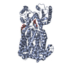
| Title | Cryo-EM structure of MmpL3 from Mycobacterium smegmatis reconstituted into peptidiscs | ||||||||||||
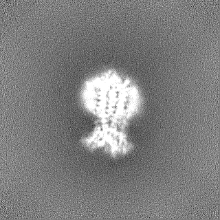
 Map data Map data | Primary map. Map from non-uniform refinement and density modification (resolution 3.23 Angstrom) | ||||||||||||
 Sample Sample |
| ||||||||||||
 Keywords Keywords | mycobacterium / trehalose monomycolate / TMM / peptidisc / MmpL3 / MEMBRANE PROTEIN | ||||||||||||
| Function / homology |  Function and homology information Function and homology informationphosphatidylethanolamine transfer activity / phosphatidylglycerol binding / trehalose transmembrane transporter activity / trehalose transport / cell wall biogenesis / mycolate cell wall layer assembly / diacylglycerol binding / cell pole / mycolic acid biosynthetic process / cell tip ...phosphatidylethanolamine transfer activity / phosphatidylglycerol binding / trehalose transmembrane transporter activity / trehalose transport / cell wall biogenesis / mycolate cell wall layer assembly / diacylglycerol binding / cell pole / mycolic acid biosynthetic process / cell tip / phospholipid transport / cell septum / phosphatidylethanolamine binding / cardiolipin binding / phosphatidylinositol binding / regulation of membrane potential / cell wall organization / response to xenobiotic stimulus / response to antibiotic / plasma membrane Similarity search - Function | ||||||||||||
| Biological species |  Mycolicibacterium smegmatis MC2 155 (bacteria) Mycolicibacterium smegmatis MC2 155 (bacteria) | ||||||||||||
| Method | single particle reconstruction / cryo EM / Resolution: 3.23 Å | ||||||||||||
 Authors Authors | Couston J / Guo Z / Wang K / Gourdon PE / Blaise M | ||||||||||||
| Funding support |  France, France,  Denmark, 3 items Denmark, 3 items
| ||||||||||||
 Citation Citation | Journal: Curr Res Struct Biol / Year: 2023 Title: Cryo-EM structure of the trehalose monomycolate transporter, MmpL3, reconstituted into peptidiscs. Authors: Julie Couston / Zongxin Guo / Kaituo Wang / Pontus Gourdon / Mickaël Blaise /  Abstract: Mycobacteria have an atypical thick and waxy cell wall. One of the major building blocks of such mycomembrane is trehalose monomycolate (TMM). TMM is a mycolic acid ester of trehalose that possesses ...Mycobacteria have an atypical thick and waxy cell wall. One of the major building blocks of such mycomembrane is trehalose monomycolate (TMM). TMM is a mycolic acid ester of trehalose that possesses long acyl chains with up to 90 carbon atoms. TMM represents an essential component of mycobacteria and is synthesized in the cytoplasm, and then flipped over the plasma membrane by a specific transporter known as MmpL3. Over the last decade, MmpL3 has emerged as an attractive drug target to combat mycobacterial infections. Recent three-dimensional structures of MmpL3 determined by X-ray crystallography and cryo-EM have increased our understanding of the TMM transport, and the mode of action of inhibiting compounds. These structures were obtained in the presence of detergent and/or in a lipidic environment. In this study, we demonstrate the possibility of obtaining a high-quality cryo-EM structure of MmpL3 without any presence of detergent through the reconstitution of the protein into peptidiscs. The structure was determined at an overall resolution of 3.2 Å and demonstrates that the overall structure of MmpL3 is preserved as compared to previous structures. Further, the study identified a new structural arrangement of the linker that fuses the two subdomains of the transmembrane domain, suggesting the feature may serve a role in the transport process. | ||||||||||||
| History |
|
- Structure visualization
| Supplemental images |
|---|

- Downloads & links
-EMDB archive
| Map data | emd_18464.map.gz | 117.7 MB | EMDB map data format | |
|---|---|---|---|---|
| Header (meta data) | emd-18464-v30.xmlemd-18464.xml | 18.8 KB 18.8 KB | Display Display | EMDB header |
| FSC (resolution estimation) | emd_18464_fsc.xml | 10.6 KB | Display | FSC data file |
| Images |  emd_18464.png emd_18464.png | 55.3 KB | ||
| Masks | emd_18464_msk_1.map | 125 MB | Mask map | |
| Filedesc metadata | emd-18464.cif.gz | 6.3 KB | ||
| Others | emd_18464_additional_1.map.gzemd_18464_half_map_1.map.gzemd_18464_half_map_2.map.gz | 62.1 MB 116 MB 116 MB | ||
| Archive directory |  http://ftp.pdbj.org/pub/emdb/structures/EMD-18464ftp://ftp.pdbj.org/pub/emdb/structures/EMD-18464 http://ftp.pdbj.org/pub/emdb/structures/EMD-18464ftp://ftp.pdbj.org/pub/emdb/structures/EMD-18464 | HTTPS FTP |
-Related structure data
| Related structure data | 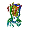 8qkkMC M: atomic model generated by this map C: citing same article ( |
|---|---|
| Similar structure data |
-Links
| EMDB pages | EMDB (EBI/PDBe) / EMDataResource |
|---|
-Map
| File | Download / File: emd_18464.map.gz / Format: CCP4 / Size: 125 MB / Type: IMAGE STORED AS FLOATING POINT NUMBER (4 BYTES) | ||||||||||||||||||||||||||||||||||||
|---|---|---|---|---|---|---|---|---|---|---|---|---|---|---|---|---|---|---|---|---|---|---|---|---|---|---|---|---|---|---|---|---|---|---|---|---|---|
| Annotation | Primary map. Map from non-uniform refinement and density modification (resolution 3.23 Angstrom) | ||||||||||||||||||||||||||||||||||||
| Projections & slices | Image control
Images are generated by Spider. | ||||||||||||||||||||||||||||||||||||
| Voxel size | X=Y=Z: 0.725 Å | ||||||||||||||||||||||||||||||||||||
| Density |
| ||||||||||||||||||||||||||||||||||||
| Symmetry | Space group: 1 | ||||||||||||||||||||||||||||||||||||
| Details | EMDB XML:
|
 Z (Sec.)
 Y (Row.)
 X (Col.)


-Supplemental data
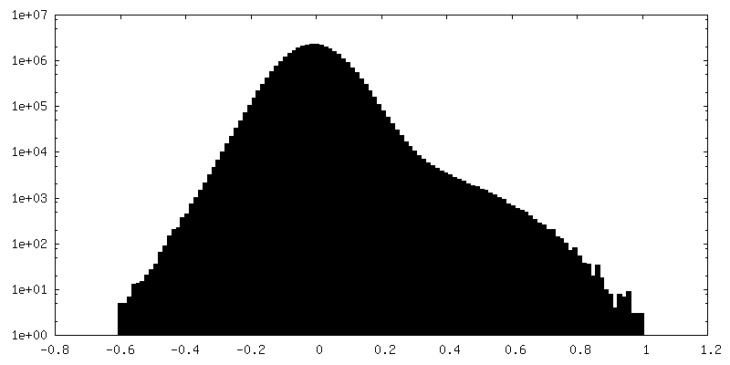
-Mask #1
| File | emd_18464_msk_1.map | ||||||||||||
|---|---|---|---|---|---|---|---|---|---|---|---|---|---|
| Projections & Slices |
| ||||||||||||
| Density Histograms |


-Additional map: map from non-uniform refinement (resolution 3.4 Angstrom)
| File | emd_18464_additional_1.map | ||||||||||||
|---|---|---|---|---|---|---|---|---|---|---|---|---|---|
| Annotation | map from non-uniform refinement (resolution 3.4 Angstrom) | ||||||||||||
| Projections & Slices |
| ||||||||||||
| Density Histograms |


-Half map: #1
| File | emd_18464_half_map_1.map | ||||||||||||
|---|---|---|---|---|---|---|---|---|---|---|---|---|---|
| Projections & Slices |
| ||||||||||||
| Density Histograms |


-Half map: #2
| File | emd_18464_half_map_2.map | ||||||||||||
|---|---|---|---|---|---|---|---|---|---|---|---|---|---|
| Projections & Slices |
| ||||||||||||
| Density Histograms |


- Sample components
-Entire : monomer structure of MmpL3
| Entire | Name: monomer structure of MmpL3 |
|---|---|
| Components |
|
-Supramolecule #1: monomer structure of MmpL3
| Supramolecule | Name: monomer structure of MmpL3 / type: complex / ID: 1 / Parent: 0 / Macromolecule list: all |
|---|---|
| Source (natural) | Organism: Mycolicibacterium smegmatis MC2 155 (bacteria) |
-Macromolecule #1: Trehalose monomycolate exporter MmpL3
| Macromolecule | Name: Trehalose monomycolate exporter MmpL3 / type: protein_or_peptide / ID: 1 / Number of copies: 1 / Enantiomer: LEVO |
|---|---|
| Source (natural) | Organism: Mycolicibacterium smegmatis MC2 155 (bacteria) |
| Molecular weight | Theoretical: 85.465344 KDa |
| Recombinant expression | Organism: |
| Sequence | String: MGFAWWGRTV YQFRYIVIGV MVALCLGGGV YGISLGNHVT QSGFYDEGSQ SVAASLIGDE VYGRDRTSHV VAILTPPDDK KVTDKAWQK KVTEELDQVV KDHEDQIVGW VGWLKAPDTT DPTVSAMKTQ DLRHTFISIP LQGDDDDEIL KNYQVVEPEL Q QVNGGDIR ...String: MGFAWWGRTV YQFRYIVIGV MVALCLGGGV YGISLGNHVT QSGFYDEGSQ SVAASLIGDE VYGRDRTSHV VAILTPPDDK KVTDKAWQK KVTEELDQVV KDHEDQIVGW VGWLKAPDTT DPTVSAMKTQ DLRHTFISIP LQGDDDDEIL KNYQVVEPEL Q QVNGGDIR LAGLNPLASE LTGTIGEDQK RAEVAAIPLV AVVLFFVFGT VIAAALPAII GGLAIAGALG IMRLVAEFTP VH FFAQPVV TLIGLGIAID YGLFIVSRFR EEIAEGYDTE AAVRRTVMTS GRTVVFSAVI IVASSVPLLL FPQGFLKSIT YAI IASVML AAILSITVLA AALAILGPRV DALGVTTLLK IPFLANWQFS RRIIDWFAEK TQKTKTREEV ERGFWGRLVN VVMK RPIAF AAPILVVMVL LIIPLGQLSL GGISEKYLPP DNAVRQSQEQ FDKLFPGFRT EPLTLVMKRE DGEPITDAQI ADMRA KALT VSGFTDPDND PEKMWKERPA NDSGSKDPSV RVIQNGLENR NDAAKKIDEL RALQPPHGIE VFVGGTPALE QDSIHS LFD KLPLMALILI VTTTVLMFLA FGSVVLPIKA ALMSALTLGS TMGILTWMFV DGHGSGLMNY TPQPLMAPMI GLIIAVI WG LSTDYEVFLV SRMVEARERG MSTAEAIRIG TATTGRLITG AALILAVVAG AFVFSDLVMM KYLAFGLLIA LLLDATII R MFLVPAVMKL LGDDCWWAPR WMKRVQEKLG LGETELPDER KRPTVRESET DQRENLYFQ UniProtKB: Trehalose monomycolate exporter MmpL3 |
-Experimental details
-Structure determination
| Method | cryo EM |
|---|---|
 Processing Processing | single particle reconstruction |
| Aggregation state | particle |
-Sample preparation
| Concentration | 3 mg/mL |
|---|---|
| Buffer | pH: 7.5 / Details: 20 mM Tris pH7.5. 150 mM NaCl |
| Sugar embedding | Material: peptidiscs |
| Grid | Model: Quantifoil R1.2/1.3 / Material: COPPER / Mesh: 300 / Support film - Material: CARBON / Support film - topology: HOLEY / Pretreatment - Type: GLOW DISCHARGE / Pretreatment - Time: 60 sec. |
| Vitrification | Cryogen name: ETHANE / Chamber humidity: 100 % / Chamber temperature: 277 K / Instrument: FEI VITROBOT MARK IV |
- Electron microscopy
| Microscope | FEI TITAN KRIOS |
|---|---|
| Image recording | Film or detector model: FEI FALCON IV (4k x 4k) / Number grids imaged: 1 / Number real images: 10004 / Average exposure time: 3.5 sec. / Average electron dose: 45.0 e/Å2 |
| Electron beam | Acceleration voltage: 300 kV / Electron source:  FIELD EMISSION GUN FIELD EMISSION GUN |
| Electron optics | Illumination mode: OTHER / Imaging mode: BRIGHT FIELD / Nominal defocus max: 2.5 µm / Nominal defocus min: 0.5 µm |
| Experimental equipment |  Model: Titan Krios / Image courtesy: FEI Company |