[English] 日本語
 Yorodumi
- PDB-8qgy: Cryo-EM structure of C-terminally truncated Apoptosis signal-regu... -
+ Open data
- Basic information
| Entry | Database: PDB / ID: 8qgy | |||||||||
|---|---|---|---|---|---|---|---|---|---|---|
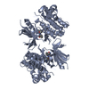
| Title | Cryo-EM structure of C-terminally truncated Apoptosis signal-regulating kinase 1 (ASK1) | |||||||||
 Components Components | Mitogen-activated protein kinase kinase kinase 5 | |||||||||
 Keywords Keywords | APOPTOSIS / ASK1 / MAP3K / MAPK signaling / thioredoxin | |||||||||
| Function / homology |  Function and homology information Function and homology informationcellular response to reactive nitrogen species / neuron intrinsic apoptotic signaling pathway in response to oxidative stress / IRE1-TRAF2-ASK1 complex / protein kinase complex / mitogen-activated protein kinase kinase kinase / programmed necrotic cell death / JUN kinase kinase kinase activity / endothelial cell apoptotic process / positive regulation of p38MAPK cascade / intrinsic apoptotic signaling pathway in response to oxidative stress ...cellular response to reactive nitrogen species / neuron intrinsic apoptotic signaling pathway in response to oxidative stress / IRE1-TRAF2-ASK1 complex / protein kinase complex / mitogen-activated protein kinase kinase kinase / programmed necrotic cell death / JUN kinase kinase kinase activity / endothelial cell apoptotic process / positive regulation of p38MAPK cascade / intrinsic apoptotic signaling pathway in response to oxidative stress / p38MAPK cascade / positive regulation of JUN kinase activity / intrinsic apoptotic signaling pathway in response to endoplasmic reticulum stress / MAP kinase kinase kinase activity / positive regulation of myoblast differentiation / positive regulation of cardiac muscle cell apoptotic process / stress-activated MAPK cascade / positive regulation of vascular associated smooth muscle cell proliferation / JNK cascade / cellular response to amino acid starvation / response to endoplasmic reticulum stress / response to ischemia / positive regulation of JNK cascade / apoptotic signaling pathway / cellular response to hydrogen peroxide / cellular response to tumor necrosis factor / cellular senescence / MAPK cascade / neuron apoptotic process / protein phosphatase binding / Oxidative Stress Induced Senescence / protein kinase activity / positive regulation of apoptotic process / protein domain specific binding / innate immune response / external side of plasma membrane / protein serine kinase activity / protein serine/threonine kinase activity / protein kinase binding / positive regulation of DNA-templated transcription / magnesium ion binding / protein homodimerization activity / protein-containing complex / ATP binding / identical protein binding / cytosol / cytoplasm Similarity search - Function | |||||||||
| Biological species |  Homo sapiens (human) Homo sapiens (human) | |||||||||
| Method | ELECTRON MICROSCOPY / single particle reconstruction / cryo EM / Resolution: 3.71 Å | |||||||||
 Authors Authors | Kosek, D. / Honzejkova, K. / Obsilova, V. / Obsil, T. | |||||||||
| Funding support |  Czech Republic, 2items Czech Republic, 2items
| |||||||||
 Citation Citation |  Journal: Elife / Year: 2024 Journal: Elife / Year: 2024Title: The cryo-EM structure of ASK1 reveals an asymmetric architecture allosterically modulated by TRX1. Authors: Karolina Honzejkova / Dalibor Kosek / Veronika Obsilova / Tomas Obsil /  Abstract: Apoptosis signal-regulating kinase 1 (ASK1) is a crucial stress sensor, directing cells toward apoptosis, differentiation, and senescence via the p38 and JNK signaling pathways. ASK1 dysregulation ...Apoptosis signal-regulating kinase 1 (ASK1) is a crucial stress sensor, directing cells toward apoptosis, differentiation, and senescence via the p38 and JNK signaling pathways. ASK1 dysregulation has been associated with cancer and inflammatory, cardiovascular, and neurodegenerative diseases, among others. However, our limited knowledge of the underlying structural mechanism of ASK1 regulation hampers our ability to target this member of the MAP3K protein family towards developing therapeutic interventions for these disorders. Nevertheless, as a multidomain Ser/Thr protein kinase, ASK1 is regulated by a complex mechanism involving dimerization and interactions with several other proteins, including thioredoxin 1 (TRX1). Thus, the present study aims at structurally characterizing ASK1 and its complex with TRX1 using several biophysical techniques. As shown by cryo-EM analysis, in a state close to its active form, ASK1 is a compact and asymmetric dimer, which enables extensive interdomain and interchain interactions. These interactions stabilize the active conformation of the ASK1 kinase domain. In turn, TRX1 functions as a negative allosteric effector of ASK1, modifying the structure of the TRX1-binding domain and changing its interaction with the tetratricopeptide repeats domain. Consequently, TRX1 reduces access to the activation segment of the kinase domain. Overall, our findings not only clarify the role of ASK1 dimerization and inter-domain contacts but also provide key mechanistic insights into its regulation, thereby highlighting the potential of ASK1 protein-protein interactions as targets for anti-inflammatory therapy. | |||||||||
| History |
|
- Structure visualization
| Structure viewer | Molecule:  Molmil Molmil Jmol/JSmol Jmol/JSmol |
|---|
- Downloads & links
- Download
| PDBx/mmCIF format |  8qgy.cif.gz 8qgy.cif.gz | 583.6 KB | Display |  PDBx/mmCIF format PDBx/mmCIF format |
|---|---|---|---|---|
| PDB format |  pdb8qgy.ent.gz pdb8qgy.ent.gz | 473.8 KB | Display |  PDB format PDB format |
| PDBx/mmJSON format |  8qgy.json.gz 8qgy.json.gz | Tree view |  PDBx/mmJSON format PDBx/mmJSON format | |
| Others |  Other downloads Other downloads |
-Validation report
| Arichive directory |  https://data.pdbj.org/pub/pdb/validation_reports/qg/8qgy https://data.pdbj.org/pub/pdb/validation_reports/qg/8qgy ftp://data.pdbj.org/pub/pdb/validation_reports/qg/8qgy ftp://data.pdbj.org/pub/pdb/validation_reports/qg/8qgy | HTTPS FTP |
|---|
-Related structure data
| Related structure data |  18396MC M: map data used to model this data C: citing same article ( |
|---|---|
| Similar structure data | Similarity search - Function & homology  F&H Search F&H Search |
- Links
Links
- Assembly
| Deposited unit | 
|
|---|---|
| 1 |
|
- Components
| #1: Protein | Mass: 101908.625 Da / Num. of mol.: 2 Source method: isolated from a genetically manipulated source Source: (gene. exp.)  Homo sapiens (human) / Gene: MAP3K5 / Production host: Homo sapiens (human) / Gene: MAP3K5 / Production host:  Has protein modification | Y | |
|---|
-Experimental details
-Experiment
| Experiment | Method: ELECTRON MICROSCOPY |
|---|---|
| EM experiment | Aggregation state: PARTICLE / 3D reconstruction method: single particle reconstruction |
- Sample preparation
| Component | Name: C-terminally truncated Apoptosis signal-regulating kinase 1 Type: COMPLEX / Entity ID: all / Source: RECOMBINANT |
|---|---|
| Molecular weight | Value: 0.2035 MDa / Experimental value: YES |
| Source (natural) | Organism:  Homo sapiens (human) Homo sapiens (human) |
| Source (recombinant) | Organism:  |
| Buffer solution | pH: 7.5 Details: 20 mM Tris-HCl 7.5 150 mM NaCl 2 mM 2-mercaptoethanol 3.8 mM CHAPSO |
| Specimen | Conc.: 0.7 mg/ml / Embedding applied: NO / Shadowing applied: NO / Staining applied: NO / Vitrification applied: YES |
| Specimen support | Details: Gatan Solarus II 955 / Grid material: GOLD / Grid mesh size: 300 divisions/in. / Grid type: UltrAuFoil R1.2/1.3 |
| Vitrification | Instrument: FEI VITROBOT MARK IV / Cryogen name: ETHANE / Humidity: 100 % / Chamber temperature: 298 K |
- Electron microscopy imaging
| Experimental equipment |  Model: Titan Krios / Image courtesy: FEI Company |
|---|---|
| Microscopy | Model: FEI TITAN KRIOS |
| Electron gun | Electron source:  FIELD EMISSION GUN / Accelerating voltage: 300 kV / Illumination mode: OTHER FIELD EMISSION GUN / Accelerating voltage: 300 kV / Illumination mode: OTHER |
| Electron lens | Mode: BRIGHT FIELD / Nominal magnification: 105000 X / Nominal defocus max: 2800 nm / Nominal defocus min: 700 nm |
| Image recording | Electron dose: 40 e/Å2 / Film or detector model: GATAN K3 BIOQUANTUM (6k x 4k) / Num. of grids imaged: 2 / Num. of real images: 5781 Details: 40 frames 2704 micrographs with 0 degree tilt, 8691 micrographs with 40 degree tilt, 5781 selected for analysis |
- Processing
| EM software |
| |||||||||||||||||||||||||||||||||||||||||||||
|---|---|---|---|---|---|---|---|---|---|---|---|---|---|---|---|---|---|---|---|---|---|---|---|---|---|---|---|---|---|---|---|---|---|---|---|---|---|---|---|---|---|---|---|---|---|---|
| CTF correction | Type: PHASE FLIPPING AND AMPLITUDE CORRECTION | |||||||||||||||||||||||||||||||||||||||||||||
| Particle selection | Num. of particles selected: 1126189 | |||||||||||||||||||||||||||||||||||||||||||||
| 3D reconstruction | Resolution: 3.71 Å / Resolution method: FSC 0.143 CUT-OFF / Num. of particles: 131110 Details: Sharpening was done using phenix.autosharpen (1.19.1-4122) Symmetry type: POINT | |||||||||||||||||||||||||||||||||||||||||||||
| Atomic model building | Protocol: FLEXIBLE FIT / Space: REAL | |||||||||||||||||||||||||||||||||||||||||||||
| Atomic model building | 3D fitting-ID: 1
| |||||||||||||||||||||||||||||||||||||||||||||
| Refine LS restraints |
|
 Movie
Movie Controller


 PDBj
PDBj

