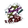
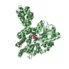
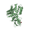
Entry Database : PDB / ID : 5ulmTitle Structure of the ASK1 central regulatory region Mitogen-activated protein kinase kinase kinase 5 Keywords / / / Function / homology Function Domain/homology Component
/ / / / / / / / / / / / / / / / / / / / / / / / / / / / / / / / / / / / / / / / / / / / / / / / / / / / / / / / / / / / / / / / / / / / / / / Biological species Homo sapiens (human)Method / / / Resolution : 2.1 Å Authors Mace, P.D. / Kumar, A. / Caradoc-Davies, T.T. Journal : Proc. Natl. Acad. Sci. U.S.A. / Year : 2017Title : Structural basis of autoregulatory scaffolding by apoptosis signal-regulating kinase 1.Authors : Weijman, J.F. / Kumar, A. / Jamieson, S.A. / King, C.M. / Caradoc-Davies, T.T. / Ledgerwood, E.C. / Murphy, J.M. / Mace, P.D. History Deposition Jan 24, 2017 Deposition site / Processing site Revision 1.0 Mar 1, 2017 Provider / Type Revision 1.1 Mar 15, 2017 Group Revision 1.2 Mar 29, 2017 Group Revision 1.3 Sep 27, 2017 Group / Data collection / Category / pdbx_audit_support / Item Revision 1.4 Nov 1, 2017 Group / Category Revision 1.5 Mar 6, 2024 Group / Database references / Refinement descriptionCategory chem_comp_atom / chem_comp_bond ... chem_comp_atom / chem_comp_bond / database_2 / struct_ncs_dom_lim Item _database_2.pdbx_DOI / _database_2.pdbx_database_accession ... _database_2.pdbx_DOI / _database_2.pdbx_database_accession / _struct_ncs_dom_lim.beg_auth_comp_id / _struct_ncs_dom_lim.beg_label_asym_id / _struct_ncs_dom_lim.beg_label_comp_id / _struct_ncs_dom_lim.beg_label_seq_id / _struct_ncs_dom_lim.end_auth_comp_id / _struct_ncs_dom_lim.end_label_asym_id / _struct_ncs_dom_lim.end_label_comp_id / _struct_ncs_dom_lim.end_label_seq_id
Show all Show less
 Movie
Movie Controller


 Open data
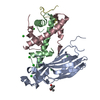
 Basic information
 Components
 Keywords
 Function and homology information
 Homo sapiens (human)
 X-RAY DIFFRACTION /
  Authors
 Citation
 Structure visualization
 Downloads & links
 Other downloads


 PDBj
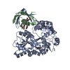
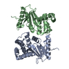
PDBj Assembly
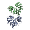


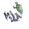
 Mass: 92.094 Da / Num. of mol.: 1 / Source method: obtained synthetically / Formula: C3H8O3
 Mass: 18.015 Da / Num. of mol.: 343 / Source method: isolated from a natural source / Formula: H2O
 Sample preparation
 / Beamline: MX2 / Wavelength: 0.979 Å
 Processing