 Movie
Movie Controller

[English] 日本語
 Yorodumi
- PDB-8qa9: Crystal structure of the RK2 plasmid encoded co-complex of the C-... -
+ Open data

- Basic information
| Entry | Database: PDB / ID: 8qa9 | ||||||||||||
|---|---|---|---|---|---|---|---|---|---|---|---|---|---|
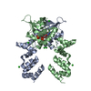
| Title | Crystal structure of the RK2 plasmid encoded co-complex of the C-terminally truncated transcriptional repressor protein KorB complexed with the partner repressor protein KorA bound to OA-DNA | ||||||||||||
 Components Components |
| ||||||||||||
 Keywords Keywords | DNA BINDING PROTEIN / multi-drug resistance / ParABS / clamp / CTP switch / DNA segregation / gene expression regulation | ||||||||||||
| Function / homology |  Function and homology information Function and homology informationchromosome segregation / chromosome / negative regulation of DNA-templated transcription / protein-containing complex / DNA binding / identical protein binding Similarity search - Function | ||||||||||||
| Biological species |  | ||||||||||||
| Method |  X-RAY DIFFRACTION / SYNCHROTRON / MOLECULAR REPLACEMENT / Resolution: 2.7 Å X-RAY DIFFRACTION / SYNCHROTRON / MOLECULAR REPLACEMENT / Resolution: 2.7 Å | ||||||||||||
 Authors Authors | McLean, T.C. / Mundy, J.E.A. / Lawson, D.M. / Le, T.B.K. | ||||||||||||
| Funding support |  United Kingdom, 3items United Kingdom, 3items
| ||||||||||||
 Citation Citation | Journal: Nat Microbiol / Year: 2025 Title: KorB switching from DNA-sliding clamp to repressor mediates long-range gene silencing in a multi-drug resistance plasmid. Authors: McLean, T.C. / Balaguer-Perez, F. / Chandanani, J. / Thomas, C.M. / Aicart-Ramos, C. / Burick, S. / Olinares, P.D.B. / Gobbato, G. / Mundy, J.E.A. / Chait, B.T. / Lawson, D.M. / Darst, S.A. ...Authors: McLean, T.C. / Balaguer-Perez, F. / Chandanani, J. / Thomas, C.M. / Aicart-Ramos, C. / Burick, S. / Olinares, P.D.B. / Gobbato, G. / Mundy, J.E.A. / Chait, B.T. / Lawson, D.M. / Darst, S.A. / Campbell, E.A. / Moreno-Herrero, F. / Le, T.B.K. | ||||||||||||
| History |
|
- Structure visualization
| Structure viewer | Molecule: MolmilJmol/JSmol |
|---|
- Downloads & links
-Download
| PDBx/mmCIF format | 8qa9.cif.gz | 295.9 KB | Display | PDBx/mmCIF format |
|---|---|---|---|---|
| PDB format | pdb8qa9.ent.gz | 238 KB | Display | PDB format |
| PDBx/mmJSON format | 8qa9.json.gz | Tree view | PDBx/mmJSON format | |
| Others |  Other downloads Other downloads |
-Validation report
| Arichive directory | https://data.pdbj.org/pub/pdb/validation_reports/qa/8qa9ftp://data.pdbj.org/pub/pdb/validation_reports/qa/8qa9 | HTTPS FTP |
|---|
-Related structure data
-Links
 PDBj
PDBj


- Assembly
| Deposited unit | 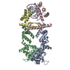
| ||||||||
|---|---|---|---|---|---|---|---|---|---|
| 1 |
| ||||||||
| Unit cell |
|
-Components
| #1: Protein | Mass: 26418.576 Da / Num. of mol.: 2 Source method: isolated from a genetically manipulated source Details: The crystallised sequence corresponds to residues 30-253 of the full 358-residue wild-type sequence appended with a C-terminal hexa-histidine tag via a short linker sequence KLAAALE. Source: (gene. exp.) #2: Protein | Mass: 12850.631 Da / Num. of mol.: 2 Source method: isolated from a genetically manipulated source Details: The crystallised sequence corresponds to the full-length 101-residue wild-type KorA protein appended with a C-terminal hexa-histidine tag via a short linker sequence KLAAALE. Source: (gene. exp.) #3: DNA chain | Mass: 4278.815 Da / Num. of mol.: 2 / Source method: obtained synthetically Details: Synthetic short double-stranded DNA sequence containing the KorA binding motif OA Source: (synth.) #4: Chemical | ChemComp-SO4 /   Mass: 96.063 Da / Num. of mol.: 5 / Source method: obtained synthetically / Formula: SO4 / Feature type: SUBJECT OF INVESTIGATION Mass: 96.063 Da / Num. of mol.: 5 / Source method: obtained synthetically / Formula: SO4 / Feature type: SUBJECT OF INVESTIGATION#5: Water | ChemComp-HOH / |  Mass: 18.015 Da / Num. of mol.: 19 / Source method: isolated from a natural source / Formula: H2O Mass: 18.015 Da / Num. of mol.: 19 / Source method: isolated from a natural source / Formula: H2OHas ligand of interest | Y | Has protein modification | N | |
|---|
-Experimental details
-Experiment
| Experiment | Method: X-RAY DIFFRACTION / Number of used crystals: 1 |
|---|
- Sample preparation
| Crystal | Density Matthews: 3.12 Å3/Da / Density % sol: 61 % |
|---|---|
| Crystal grow | Temperature: 293 K / Method: vapor diffusion, sitting drop / pH: 6 / Details: NULL |
-Data collection
| Diffraction | Mean temperature: 100 K / Serial crystal experiment: N |
|---|---|
| Diffraction source | Source: SYNCHROTRON / Site: Diamond / Beamline: I03 / Wavelength: 0.9763 Å |
| Detector | Type: DECTRIS EIGER2 XE 16M / Detector: PIXEL / Date: Jul 14, 2022 |
| Radiation | Protocol: SINGLE WAVELENGTH / Monochromatic (M) / Laue (L): M / Scattering type: x-ray |
| Radiation wavelength | Wavelength: 0.9763 Å / Relative weight: 1 |
| Reflection | Resolution: 2.7→82.65 Å / Num. obs: 29438 / % possible obs: 100 % / Redundancy: 12.4 % / Biso Wilson estimate: 58.1 Å2 / CC1/2: 0.993 / Rmerge(I) obs: 0.13 / Rpim(I) all: 0.039 / Rrim(I) all: 0.135 / Χ2: 0.59 / Net I/σ(I): 14.1 |
| Reflection shell | Resolution: 2.7→2.83 Å / % possible obs: 100 % / Redundancy: 11.6 % / Rmerge(I) obs: 1.533 / Num. measured all: 45107 / Num. unique obs: 3879 / CC1/2: 0.677 / Rpim(I) all: 0.473 / Rrim(I) all: 1.606 / Χ2: 0.21 / Net I/σ(I) obs: 1 |
- Processing
| Software |
| ||||||||||||||||||||||||||||||||||||||||||||||||||||||||||||||||||||||||||||||||||||||||||||||||||||||||||||||||||||||||||||||||||||||||||||||||||||||||||||||||||||||||||||||||||||||
|---|---|---|---|---|---|---|---|---|---|---|---|---|---|---|---|---|---|---|---|---|---|---|---|---|---|---|---|---|---|---|---|---|---|---|---|---|---|---|---|---|---|---|---|---|---|---|---|---|---|---|---|---|---|---|---|---|---|---|---|---|---|---|---|---|---|---|---|---|---|---|---|---|---|---|---|---|---|---|---|---|---|---|---|---|---|---|---|---|---|---|---|---|---|---|---|---|---|---|---|---|---|---|---|---|---|---|---|---|---|---|---|---|---|---|---|---|---|---|---|---|---|---|---|---|---|---|---|---|---|---|---|---|---|---|---|---|---|---|---|---|---|---|---|---|---|---|---|---|---|---|---|---|---|---|---|---|---|---|---|---|---|---|---|---|---|---|---|---|---|---|---|---|---|---|---|---|---|---|---|---|---|---|---|
| Refinement | Method to determine structure: MOLECULAR REPLACEMENT / Resolution: 2.7→82.65 Å / Cor.coef. Fo:Fc: 0.95 / Cor.coef. Fo:Fc free: 0.83 / SU B: 30.2 / SU ML: 0.268 / Cross valid method: THROUGHOUT / ESU R: 0.521 / ESU R Free: 0.31 / Stereochemistry target values: MAXIMUM LIKELIHOOD / Details: HYDROGENS HAVE BEEN ADDED IN THE RIDING POSITIONS
| ||||||||||||||||||||||||||||||||||||||||||||||||||||||||||||||||||||||||||||||||||||||||||||||||||||||||||||||||||||||||||||||||||||||||||||||||||||||||||||||||||||||||||||||||||||||
| Solvent computation | Ion probe radii: 0.8 Å / Shrinkage radii: 0.8 Å / VDW probe radii: 1.2 Å / Solvent model: MASK | ||||||||||||||||||||||||||||||||||||||||||||||||||||||||||||||||||||||||||||||||||||||||||||||||||||||||||||||||||||||||||||||||||||||||||||||||||||||||||||||||||||||||||||||||||||||
| Displacement parameters | Biso mean: 76.613 Å2
| ||||||||||||||||||||||||||||||||||||||||||||||||||||||||||||||||||||||||||||||||||||||||||||||||||||||||||||||||||||||||||||||||||||||||||||||||||||||||||||||||||||||||||||||||||||||
| Refinement step | Cycle: 1 / Resolution: 2.7→82.65 Å
| ||||||||||||||||||||||||||||||||||||||||||||||||||||||||||||||||||||||||||||||||||||||||||||||||||||||||||||||||||||||||||||||||||||||||||||||||||||||||||||||||||||||||||||||||||||||
| Refine LS restraints |
|