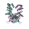
[English] 日本語
Yorodumi
- PDB-8qa8: Crystal structure of the C-terminally truncated transcriptional r... -

+
Open data


ID or keywords:

Loading...

-
Basic information

Entry
Database: PDB / ID: 8qa8
TitleCrystal structure of the C-terminally truncated transcriptional repressor protein KorB from the RK2 plasmid complexed with CTP-gamma-S
ComponentsTranscriptional repressor protein KorB
KeywordsDNA BINDING PROTEIN / multi-drug resistance / ParABS / clamp / CTP switch / DNA segregation / gene expression regulation
Function / homology
Function and homology information


negative regulation of DNA-templated transcription / protein-containing complex / DNA binding / identical protein binding
Similarity search - Function
KorB, C-terminal / Repressor KorB domain / KorB, C-terminal domain superfamily / KorB DNA-binding domain / KorB C-terminal beta-barrel domain / KorB domain / ParB/RepB/Spo0J partition protein / ParB/Sulfiredoxin domain / ParB/Sulfiredoxin / ParB-like nuclease domain ...KorB, C-terminal / Repressor KorB domain / KorB, C-terminal domain superfamily / KorB DNA-binding domain / KorB C-terminal beta-barrel domain / KorB domain / ParB/RepB/Spo0J partition protein / ParB/Sulfiredoxin domain / ParB/Sulfiredoxin / ParB-like nuclease domain / ParB/Sulfiredoxin superfamily / Transcriptional repressor, C-terminal
Similarity search - Domain/homology
CYTIDINE-5'-TRIPHOSPHATE / Transcriptional repressor protein KorB
Similarity search - Component
Biological speciesEscherichia coli (E. coli)
MethodX-RAY DIFFRACTION / SYNCHROTRON / MOLECULAR REPLACEMENT / Resolution: 2.3 Å
AuthorsMcLean, T.C. / Mundy, J.E.A. / Lawson, D.M. / Le, T.B.K.
Funding support United Kingdom, 3items
OrganizationGrant numberCountry
Biotechnology and Biological Sciences Research Council (BBSRC)BB/X01097X/1 United Kingdom
Wellcome Trust221776/Z/20/Z United Kingdom
Royal SocietyURF/R/201020 United Kingdom
CitationJournal: To Be Published
Title: Crystal structure of the C-terminally truncated transcriptional repressor protein KorB from the RK2 plasmid complexed with CTP-gamma-S
Authors: McLean, T.C. / Mundy, J.E.A. / Lawson, D.M. / Le, T.B.K.
History
DepositionAug 22, 2023Deposition site: PDBE / Processing site: PDBE
Revision 1.0Feb 21, 2024Provider: repository / Type: Initial release

-
Structure visualization

Structure viewerMolecule:
MolmilJmol/JSmol

Downloads & links

-
Assembly

Deposited unit
A: Transcriptional repressor protein KorB
B: Transcriptional repressor protein KorB
C: Transcriptional repressor protein KorB
D: Transcriptional repressor protein KorB
E: Transcriptional repressor protein KorB
F: Transcriptional repressor protein KorB
hetero molecules


Theoretical massNumber of molelcules
Total (without water)162,19034
Polymers158,5116
Non-polymers3,67928
Water3,675204
1
A: Transcriptional repressor protein KorB
B: Transcriptional repressor protein KorB
hetero molecules


Theoretical massNumber of molelcules
Total (without water)54,08712
Polymers52,8372
Non-polymers1,25010
Water362
TypeNameSymmetry operationNumber
identity operation1_555x,y,z1
Buried area7000 Å2
ΔGint-78 kcal/mol
Surface area20480 Å2
MethodPISA
2
C: Transcriptional repressor protein KorB
D: Transcriptional repressor protein KorB
hetero molecules


Theoretical massNumber of molelcules
Total (without water)54,05211
Polymers52,8372
Non-polymers1,2149
Water362
TypeNameSymmetry operationNumber
identity operation1_555x,y,z1
Buried area7200 Å2
ΔGint-70 kcal/mol
Surface area20240 Å2
MethodPISA
3
E: Transcriptional repressor protein KorB
F: Transcriptional repressor protein KorB
hetero molecules


Theoretical massNumber of molelcules
Total (without water)54,05211
Polymers52,8372
Non-polymers1,2149
Water362
TypeNameSymmetry operationNumber
identity operation1_555x,y,z1
Buried area6860 Å2
ΔGint-69 kcal/mol
Surface area19950 Å2
MethodPISA
Unit cell
Length a, b, c (Å)58.686, 152.710, 198.274
Angle α, β, γ (deg.)90.00, 90.00, 90.00
Int Tables number19
Space group name H-MP212121

-
Components

#1: Protein
Transcriptional repressor protein KorB


Mass: 26418.576 Da / Num. of mol.: 6
Source method: isolated from a genetically manipulated source
Details: The crystallised sequence corresponds to residues 30-253 of the full 358-residue wild-type sequence appended with a C-terminal hexa-histidine tag via a short linker sequence KLAAALE.
Source: (gene. exp.) Escherichia coli (E. coli) / Gene: korB / Plasmid: pET21b / Production host: Escherichia coli BL21(DE3) (bacteria) / References: UniProt: P07674
#2: Chemical
ChemComp-CTP / CYTIDINE-5'-TRIPHOSPHATE / Cytidine triphosphate


Mass: 483.156 Da / Num. of mol.: 6 / Source method: obtained synthetically / Formula: C9H16N3O14P3 / Feature type: SUBJECT OF INVESTIGATION
#3: Chemical...
ChemComp-CL / CHLORIDE ION / Chloride


Mass: 35.453 Da / Num. of mol.: 22 / Source method: isolated from a natural source / Formula: Cl
#4: Water ChemComp-HOH / water / Water


Mass: 18.015 Da / Num. of mol.: 204 / Source method: isolated from a natural source / Formula: H2O
Has ligand of interestY

-
Experimental details

-
Experiment

ExperimentMethod: X-RAY DIFFRACTION / Number of used crystals: 1

-
Sample preparation

CrystalDensity Matthews: 2.89 Å3/Da / Density % sol: 57 %
Crystal growTemperature: 293 K / Method: vapor diffusion, sitting drop / Details: NULL

-
Data collection

DiffractionMean temperature: 100 K / Serial crystal experiment: N
Diffraction sourceSource: SYNCHROTRON / Site: Diamond / Beamline: I04-1 / Wavelength: 0.9179 Å
DetectorType: DECTRIS EIGER2 XE 9M / Detector: PIXEL / Date: Mar 2, 2022
RadiationProtocol: SINGLE WAVELENGTH / Monochromatic (M) / Laue (L): M / Scattering type: x-ray
Radiation wavelengthWavelength: 0.9179 Å / Relative weight: 1
ReflectionResolution: 2.3→198.27 Å / Num. obs: 80304 / % possible obs: 100 % / Redundancy: 13.3 % / CC1/2: 0.997 / Rmerge(I) obs: 0.236 / Rpim(I) all: 0.067 / Rrim(I) all: 0.245 / Χ2: 0.43 / Net I/σ(I): 7.9 / Num. measured all: 1069894
Reflection shellResolution: 2.3→2.35 Å / % possible obs: 100 % / Redundancy: 11.1 % / Rmerge(I) obs: 1.887 / Num. measured all: 50482 / Num. unique obs: 4563 / CC1/2: 0.718 / Rpim(I) all: 0.594 / Rrim(I) all: 1.979 / Χ2: 0.18 / Net I/σ(I) obs: 0.7

-
Processing

Software
NameVersionClassification
REFMAC5.8.0403refinement
Aimlessdata scaling
DIALSdata reduction
PHASERphasing
RefinementMethod to determine structure: MOLECULAR REPLACEMENT / Resolution: 2.3→121.28 Å / Cor.coef. Fo:Fc: 0.934 / Cor.coef. Fo:Fc free: 0.922 / SU B: 20.803 / SU ML: 0.225 / Cross valid method: THROUGHOUT / ESU R: 0.279 / ESU R Free: 0.217 / Stereochemistry target values: MAXIMUM LIKELIHOOD / Details: HYDROGENS HAVE BEEN ADDED IN THE RIDING POSITIONS
RfactorNum. reflection% reflectionSelection details
Rfree0.25619 4000 5 %RANDOM
Rwork0.2274 ---
obs0.22881 76214 100 %-
Solvent computationIon probe radii: 0.8 Å / Shrinkage radii: 0.8 Å / VDW probe radii: 1.2 Å / Solvent model: MASK
Displacement parametersBiso mean: 55.721 Å2
Baniso -1Baniso -2Baniso -3
1-0.54 Å20 Å20 Å2
2---0.97 Å20 Å2
3---0.43 Å2
Refinement stepCycle: 1 / Resolution: 2.3→121.28 Å
ProteinNucleic acidLigandSolventTotal
Num. atoms9414 0 196 204 9814
Refine LS restraints
Refine-IDTypeDev idealDev ideal targetNumber
X-RAY DIFFRACTIONr_bond_refined_d0.0090.0129958
X-RAY DIFFRACTIONr_bond_other_d0.0010.0169384
X-RAY DIFFRACTIONr_angle_refined_deg1.4411.66113548
X-RAY DIFFRACTIONr_angle_other_deg0.4691.57921651
X-RAY DIFFRACTIONr_dihedral_angle_1_deg5.8451229
X-RAY DIFFRACTIONr_dihedral_angle_2_deg9.797587
X-RAY DIFFRACTIONr_dihedral_angle_3_deg16.229101708
X-RAY DIFFRACTIONr_dihedral_angle_4_deg
X-RAY DIFFRACTIONr_chiral_restr0.0710.21495
X-RAY DIFFRACTIONr_gen_planes_refined0.0070.0211864
X-RAY DIFFRACTIONr_gen_planes_other0.0010.022223
X-RAY DIFFRACTIONr_nbd_refined
X-RAY DIFFRACTIONr_nbd_other
X-RAY DIFFRACTIONr_nbtor_refined
X-RAY DIFFRACTIONr_nbtor_other
X-RAY DIFFRACTIONr_xyhbond_nbd_refined
X-RAY DIFFRACTIONr_xyhbond_nbd_other
X-RAY DIFFRACTIONr_metal_ion_refined
X-RAY DIFFRACTIONr_metal_ion_other
X-RAY DIFFRACTIONr_symmetry_vdw_refined
X-RAY DIFFRACTIONr_symmetry_vdw_other
X-RAY DIFFRACTIONr_symmetry_hbond_refined
X-RAY DIFFRACTIONr_symmetry_hbond_other
X-RAY DIFFRACTIONr_symmetry_metal_ion_refined
X-RAY DIFFRACTIONr_symmetry_metal_ion_other
X-RAY DIFFRACTIONr_mcbond_it1.8322.0894883
X-RAY DIFFRACTIONr_mcbond_other1.8312.094883
X-RAY DIFFRACTIONr_mcangle_it2.9583.7436123
X-RAY DIFFRACTIONr_mcangle_other2.9583.7436124
X-RAY DIFFRACTIONr_scbond_it2.5522.2865075
X-RAY DIFFRACTIONr_scbond_other2.5422.2865064
X-RAY DIFFRACTIONr_scangle_it
X-RAY DIFFRACTIONr_scangle_other4.1454.097426
X-RAY DIFFRACTIONr_long_range_B_refined6.15619.3310842
X-RAY DIFFRACTIONr_long_range_B_other6.15419.310825
X-RAY DIFFRACTIONr_rigid_bond_restr
X-RAY DIFFRACTIONr_sphericity_free
X-RAY DIFFRACTIONr_sphericity_bonded
LS refinement shellResolution: 2.3→2.36 Å / Total num. of bins used: 20
RfactorNum. reflection% reflection
Rfree0.348 269 -
Rwork0.349 5607 -
obs--100 %
Refinement TLS params.

Method: refined / Refine-ID: X-RAY DIFFRACTION

IDL112)L122)L132)L222)L232)L332)S11 (Å °)S12 (Å °)S13 (Å °)S21 (Å °)S22 (Å °)S23 (Å °)S31 (Å °)S32 (Å °)S33 (Å °)T112)T122)T132)T222)T232)T332)Origin x (Å)Origin y (Å)Origin z (Å)
13.07521.3186-0.08713.0082-0.80171.54960.03390.10.33130.0814-0.04810.1075-0.2313-0.0160.01420.03810.0392-0.00120.5064-0.0210.040528.8652-8.1322.4792
23.2651-1.0706-1.32131.89250.73092.97620.0231-0.1420.28940.01480.0431-0.112-0.290.0756-0.06610.0384-0.0404-0.00880.4612-0.0320.064827.3244-12.936815.3856
31.94690.0584-0.4092.1261-0.15233.2863-0.00290.1063-0.0563-0.1470.0873-0.00120.14310.0485-0.08440.0172-0.01260.00010.46840.05580.226275.56963.0234-23.6505
43.54590.8731-0.35822.3109-0.67912.32590.02120.21220.2254-0.25530.0240.2163-0.0607-0.1087-0.04520.05730.07470.0210.62310.08610.147861.7566.5759-22.621
52.33160.7216-0.41313.1404-0.56093.53270.00220.0145-0.36960.0941-0.03740.03730.3095-0.09590.03520.04830.0254-0.02820.4916-0.09910.147939.2539-21.432443.139
63.5553-0.7026-1.48931.67010.55454.0219-0.0719-0.0345-0.35650.0004-0.0258-0.140.31760.11030.09770.0596-0.0202-0.0330.45580.00850.153553.0411-17.36344.2564
Refinement TLS group
IDRefine-IDRefine TLS-IDAuth asym-IDAuth seq-ID
1X-RAY DIFFRACTION1A52 - 252
2X-RAY DIFFRACTION2B52 - 253
3X-RAY DIFFRACTION3C52 - 251
4X-RAY DIFFRACTION4D52 - 252
5X-RAY DIFFRACTION5E52 - 251
6X-RAY DIFFRACTION6F52 - 248

+
About Yorodumi

-
News

-
Feb 9, 2022. New format data for meta-information of EMDB entries

New format data for meta-information of EMDB entries

  • Version 3 of the EMDB header file is now the official format.
  • The previous official version 1.9 will be removed from the archive.

Related info.:EMDB header

External links:wwPDB to switch to version 3 of the EMDB data model

-
Aug 12, 2020. Covid-19 info

Covid-19 info

URL: https://pdbj.org/emnavi/covid19.php

New page: Covid-19 featured information page in EM Navigator.

Related info.:Covid-19 info / Mar 5, 2020. Novel coronavirus structure data

+
Mar 5, 2020. Novel coronavirus structure data

Novel coronavirus structure data

Related info.:Yorodumi Speices / Aug 12, 2020. Covid-19 info

External links:COVID-19 featured content - PDBj / Molecule of the Month (242):Coronavirus Proteases

+
Jan 31, 2019. EMDB accession codes are about to change! (news from PDBe EMDB page)

EMDB accession codes are about to change! (news from PDBe EMDB page)

  • The allocation of 4 digits for EMDB accession codes will soon come to an end. Whilst these codes will remain in use, new EMDB accession codes will include an additional digit and will expand incrementally as the available range of codes is exhausted. The current 4-digit format prefixed with “EMD-” (i.e. EMD-XXXX) will advance to a 5-digit format (i.e. EMD-XXXXX), and so on. It is currently estimated that the 4-digit codes will be depleted around Spring 2019, at which point the 5-digit format will come into force.
  • The EM Navigator/Yorodumi systems omit the EMD- prefix.

Related info.:Q: What is EMD? / ID/Accession-code notation in Yorodumi/EM Navigator

External links:EMDB Accession Codes are Changing Soon! / Contact to PDBj

+
Jul 12, 2017. Major update of PDB

Major update of PDB

  • wwPDB released updated PDB data conforming to the new PDBx/mmCIF dictionary.
  • This is a major update changing the version number from 4 to 5, and with Remediation, in which all the entries are updated.
  • In this update, many items about electron microscopy experimental information are reorganized (e.g. em_software).
  • Now, EM Navigator and Yorodumi are based on the updated data.

External links:wwPDB Remediation / Enriched Model Files Conforming to OneDep Data Standards Now Available in the PDB FTP Archive

-
Yorodumi

Thousand views of thousand structures

  • Yorodumi is a browser for structure data from EMDB, PDB, SASBDB, etc.
  • This page is also the successor to EM Navigator detail page, and also detail information page/front-end page for Omokage search.
  • The word "yorodu" (or yorozu) is an old Japanese word meaning "ten thousand". "mi" (miru) is to see.

Related info.:EMDB / PDB / SASBDB / Comparison of 3 databanks / Yorodumi Search / Aug 31, 2016. New EM Navigator & Yorodumi / Yorodumi Papers / Jmol/JSmol / Function and homology information / Changes in new EM Navigator and Yorodumi

Read more