 Movie
Movie Controller

[English] 日本語
 Yorodumi
- PDB-8q9n: Crystal Structure of the MADS-box/MEF2 Domain of MEF2D bound to d... -
+ Open data

- Basic information
| Entry | Database: PDB / ID: 8q9n | ||||||
|---|---|---|---|---|---|---|---|
| Title | Crystal Structure of the MADS-box/MEF2 Domain of MEF2D bound to dsDNA and MITR deacetylase binding motif mutant L151V. | ||||||
 Components Components |
| ||||||
 Keywords Keywords | TRANSCRIPTION / transcriptional repressor | ||||||
| Function / homology |  Function and homology information Function and homology informationanimal organ development / RNA polymerase II transcription regulatory region sequence-specific DNA binding / nervous system development / DNA-binding transcription factor activity, RNA polymerase II-specific / protein dimerization activity / positive regulation of transcription by RNA polymerase II / nucleus Similarity search - Function | ||||||
| Biological species |  Homo sapiens (human) Homo sapiens (human) | ||||||
| Method |  X-RAY DIFFRACTION / SYNCHROTRON / MOLECULAR REPLACEMENT / Resolution: 1.51 Å X-RAY DIFFRACTION / SYNCHROTRON / MOLECULAR REPLACEMENT / Resolution: 1.51 Å | ||||||
 Authors Authors | Chinellato, M. / Carli, A. / Perin, S. / Mazzocato, Y. / Biondi, B. / Di Giorgio, E. / Brancolini, C. / Angelini, A. / Cendron, L. | ||||||
| Funding support |  Italy, 1items Italy, 1items
| ||||||
 Citation Citation | Journal: J.Mol.Biol. / Year: 2024 Title: Folding of Class IIa HDAC Derived Peptides into alpha-helices Upon Binding to Myocyte Enhancer Factor-2 in Complex with DNA. Authors: Chinellato, M. / Perin, S. / Carli, A. / Lastella, L. / Biondi, B. / Borsato, G. / Di Giorgio, E. / Brancolini, C. / Cendron, L. / Angelini, A. | ||||||
| History |
|
- Structure visualization
| Structure viewer | Molecule: MolmilJmol/JSmol |
|---|
- Downloads & links
-Download
| PDBx/mmCIF format | 8q9n.cif.gz | 77.4 KB | Display | PDBx/mmCIF format |
|---|---|---|---|---|
| PDB format | pdb8q9n.ent.gz | 54 KB | Display | PDB format |
| PDBx/mmJSON format | 8q9n.json.gz | Tree view | PDBx/mmJSON format | |
| Others |  Other downloads Other downloads |
-Validation report
| Arichive directory | https://data.pdbj.org/pub/pdb/validation_reports/q9/8q9nftp://data.pdbj.org/pub/pdb/validation_reports/q9/8q9n | HTTPS FTP |
|---|
-Related structure data


-Links
 PDBj
PDBj


- Assembly
| Deposited unit | 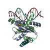
| ||||||||
|---|---|---|---|---|---|---|---|---|---|
| 1 |
| ||||||||
| Unit cell |
|
-Components
-DNA chain , 2 types, 2 molecules KL
| #2: DNA chain | Mass: 4286.842 Da / Num. of mol.: 1 / Source method: obtained synthetically / Source: (synth.) Homo sapiens (human) |
|---|---|
| #3: DNA chain | Mass: 4268.813 Da / Num. of mol.: 1 / Source method: obtained synthetically / Source: (synth.) Homo sapiens (human) |
-Protein / Protein/peptide , 2 types, 3 molecules ABX
| #1: Protein | Mass: 11254.035 Da / Num. of mol.: 2 Source method: isolated from a genetically manipulated source Details: Missing residues are not visible in the electron density maps Source: (gene. exp.) Homo sapiens (human) / Gene: MEF2D / Production host:  #4: Protein/peptide | | Mass: 2079.375 Da / Num. of mol.: 1 / Source method: obtained synthetically Details: Missing residues are not visible in the electron density maps Source: (synth.) Homo sapiens (human) |
|---|
-Non-polymers , 2 types, 150 molecules 

| #5: Chemical | ChemComp-DMS /  Mass: 78.133 Da / Num. of mol.: 1 / Source method: obtained synthetically / Formula: C2H6OS / Comment: DMSO, precipitant*YM Mass: 78.133 Da / Num. of mol.: 1 / Source method: obtained synthetically / Formula: C2H6OS / Comment: DMSO, precipitant*YM |
|---|---|
| #6: Water | ChemComp-HOH / Mass: 18.015 Da / Num. of mol.: 149 / Source method: isolated from a natural source / Formula: H2O |
-Details
| Has ligand of interest | N |
|---|---|
| Has protein modification | Y |
-Experimental details
-Experiment
| Experiment | Method: X-RAY DIFFRACTION / Number of used crystals: 1 |
|---|
- Sample preparation
| Crystal | Density Matthews: 1.79 Å3/Da / Density % sol: 31.39 % |
|---|---|
| Crystal grow | Temperature: 293 K / Method: vapor diffusion, sitting drop Details: 200 mM sodium formate, 100 mM Bis-Tris propane pH 6.5 and 20% w/v polyethylene glycol 3350 (PEG 3350) |
-Data collection
| Diffraction | Mean temperature: 100 K / Serial crystal experiment: N |
|---|---|
| Diffraction source | Source: SYNCHROTRON / Site: ESRF  / Beamline: ID23-2 / Wavelength: 0.8731 Å / Beamline: ID23-2 / Wavelength: 0.8731 Å |
| Detector | Type: DECTRIS PILATUS 2M / Detector: PIXEL / Date: Nov 23, 2020 |
| Radiation | Protocol: SINGLE WAVELENGTH / Monochromatic (M) / Laue (L): M / Scattering type: x-ray |
| Radiation wavelength | Wavelength: 0.8731 Å / Relative weight: 1 |
| Reflection | Resolution: 1.51→45.8 Å / Num. obs: 36328 / % possible obs: 100 % / Redundancy: 7 % / CC1/2: 0.999 / Rmerge(I) obs: 0.076 / Net I/σ(I): 12.7 |
| Reflection shell | Resolution: 1.6→1.63 Å / Rmerge(I) obs: 0.861 / Mean I/σ(I) obs: 1.9 / Num. unique obs: 1562 / CC1/2: 0.752 |
- Processing
| Software |
| ||||||||||||||||||||
|---|---|---|---|---|---|---|---|---|---|---|---|---|---|---|---|---|---|---|---|---|---|
| Refinement | Method to determine structure: MOLECULAR REPLACEMENT / Resolution: 1.51→45.76 Å / SU B: 1.857 / SU ML: 0.067 / Cross valid method: THROUGHOUT / ESU R: 0.089 / ESU R Free: 0.096 / Details: HYDROGENS HAVE BEEN USED IF PRESENT IN THE INPUT
| ||||||||||||||||||||
| Displacement parameters | Biso mean: 26.105 Å2
| ||||||||||||||||||||
| Refinement step | Cycle: LAST / Resolution: 1.51→45.76 Å
|
