[English] 日本語
Yorodumi
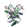
- PDB-8c84: Crystal structure of MADS-box/MEF2D N-terminal domain complex -

+
Open data


ID or keywords:

Loading...

-
Basic information

Entry
Database: PDB / ID: 8c84
TitleCrystal structure of MADS-box/MEF2D N-terminal domain complex
Components
  • DNA (5'-D(P*AP*AP*CP*TP*AP*TP*TP*TP*AP*TP*AP*AP*GP*A)-3')
  • DNA (5'-D(P*TP*CP*TP*TP*AP*TP*AP*AP*AP*TP*AP*GP*TP*T)-3')
  • MEF2D protein
KeywordsTRANSCRIPTION / transcriptional regulator
Function / homology
Function and homology information


histone deacetylase binding / heart development / cell differentiation / protein dimerization activity / DNA-binding transcription factor activity, RNA polymerase II-specific / RNA polymerase II cis-regulatory region sequence-specific DNA binding / positive regulation of transcription by RNA polymerase II / nucleus
Similarity search - Function
Holliday junction regulator protein family C-terminal / Holliday junction regulator protein family C-terminal repeat / MADS MEF2-like / Transcription factor, MADS-box / Transcription factor, MADS-box superfamily / SRF-type transcription factor (DNA-binding and dimerisation domain) / MADS-box domain signature. / MADS-box domain profile. / MADS
Similarity search - Domain/homology
DNA / DNA (> 10) / MEF2D protein
Similarity search - Component
Biological speciesHomo sapiens (human)
synthetic construct (others)
MethodX-RAY DIFFRACTION / SYNCHROTRON / MOLECULAR REPLACEMENT / Resolution: 1.9 Å
AuthorsChinellato, M. / Carli, A. / Perin, S. / Mazzoccato, Y. / Di Giorgio, E. / Brancolini, C. / Angelini, A. / Cendron, L.
Funding support Italy, 1items
OrganizationGrant numberCountry
Italian Ministry of EducationC94I19001550001 Italy
CitationJournal: J.Mol.Biol. / Year: 2024
Title: Folding of Class IIa HDAC Derived Peptides into alpha-helices Upon Binding to Myocyte Enhancer Factor-2 in Complex with DNA.
Authors: Chinellato, M. / Perin, S. / Carli, A. / Lastella, L. / Biondi, B. / Borsato, G. / Di Giorgio, E. / Brancolini, C. / Cendron, L. / Angelini, A.
History
DepositionJan 18, 2023Deposition site: PDBE / Processing site: PDBE
Revision 1.0Jan 31, 2024Provider: repository / Type: Initial release
Revision 1.1Apr 17, 2024Group: Database references / Category: citation / citation_author
Item: _citation.country / _citation.journal_abbrev ..._citation.country / _citation.journal_abbrev / _citation.journal_id_ASTM / _citation.journal_id_CSD / _citation.journal_id_ISSN / _citation.journal_volume / _citation.page_first / _citation.page_last / _citation.pdbx_database_id_DOI / _citation.pdbx_database_id_PubMed / _citation.title / _citation.year

-
Structure visualization

Structure viewerMolecule:
MolmilJmol/JSmol

Downloads & links

-
Assembly

Deposited unit
A: MEF2D protein
B: MEF2D protein
K: DNA (5'-D(P*AP*AP*CP*TP*AP*TP*TP*TP*AP*TP*AP*AP*GP*A)-3')
L: DNA (5'-D(P*TP*CP*TP*TP*AP*TP*AP*AP*AP*TP*AP*GP*TP*T)-3')


Theoretical massNumber of molelcules
Total (without water)30,6874
Polymers30,6874
Non-polymers00
Water1,31573
1


  • Idetical with deposited unit
  • defined by author&software
  • Evidence: gel filtration
TypeNameSymmetry operationNumber
identity operation1_555x,y,z1
Buried area10660 Å2
ΔGint-62 kcal/mol
Surface area13670 Å2
MethodPISA
Unit cell
Length a, b, c (Å)53.033, 56.364, 75.596
Angle α, β, γ (deg.)90.00, 90.00, 90.00
Int Tables number19
Space group name H-MP212121

-
Components

#1: Protein MEF2D protein / Myocyte-specific enhancer factor 2D


Mass: 11065.788 Da / Num. of mol.: 2
Source method: isolated from a genetically manipulated source
Details: missing residues are not visible in the electron density maps
Source: (gene. exp.) Homo sapiens (human) / Gene: MEF2D / Production host: Escherichia coli BL21(DE3) (bacteria) / References: UniProt: Q05BX2
#2: DNA chain DNA (5'-D(P*AP*AP*CP*TP*AP*TP*TP*TP*AP*TP*AP*AP*GP*A)-3')


Mass: 4286.842 Da / Num. of mol.: 1 / Source method: obtained synthetically / Source: (synth.) synthetic construct (others)
#3: DNA chain DNA (5'-D(P*TP*CP*TP*TP*AP*TP*AP*AP*AP*TP*AP*GP*TP*T)-3')


Mass: 4268.813 Da / Num. of mol.: 1 / Source method: obtained synthetically / Source: (synth.) synthetic construct (others)
#4: Water ChemComp-HOH / water


Mass: 18.015 Da / Num. of mol.: 73 / Source method: isolated from a natural source / Formula: H2O

-
Experimental details

-
Experiment

ExperimentMethod: X-RAY DIFFRACTION / Number of used crystals: 1

-
Sample preparation

CrystalDensity Matthews: 1.84 Å3/Da / Density % sol: 33.32 %
Crystal growTemperature: 293 K / Method: vapor diffusion, sitting drop
Details: 200 mM sodium acetate trihydrate, 100 mM Bis-Tris propane pH 6.5 and 20% w/v polyethylene glycol 3350 (PEG 3350)

-
Data collection

DiffractionMean temperature: 100 K / Serial crystal experiment: N
Diffraction sourceSource: SYNCHROTRON / Site: ESRF / Beamline: ID30B / Wavelength: 0.9724 Å
DetectorType: DECTRIS EIGER2 X 16M / Detector: PIXEL / Date: Dec 9, 2020
RadiationProtocol: SINGLE WAVELENGTH / Monochromatic (M) / Laue (L): M / Scattering type: x-ray
Radiation wavelengthWavelength: 0.9724 Å / Relative weight: 1
ReflectionResolution: 1.9→45.19 Å / Num. obs: 18562 / % possible obs: 99.8 % / Redundancy: 8.1 % / CC1/2: 0.999 / Rmerge(I) obs: 0.07 / Net I/σ(I): 15.1
Reflection shellResolution: 1.9→1.94 Å / Rmerge(I) obs: 0.893 / Num. unique obs: 1182 / CC1/2: 0.791

-
Processing

Software
NameVersionClassification
REFMAC5.8.0403refinement
PHENIX1.18.2refinement
xia2data reduction
Aimlessdata scaling
MOLREPphasing
RefinementMethod to determine structure: MOLECULAR REPLACEMENT / Resolution: 1.9→45.19 Å / Cor.coef. Fo:Fc: 0.961 / Cor.coef. Fo:Fc free: 0.948 / SU B: 4.414 / SU ML: 0.126 / Cross valid method: THROUGHOUT / ESU R: 0.196 / ESU R Free: 0.171 / Stereochemistry target values: MAXIMUM LIKELIHOOD
RfactorNum. reflection% reflectionSelection details
Rfree0.25118 952 5.2 %RANDOM
Rwork0.20377 ---
obs0.20619 18412 99.8 %-
Solvent computationIon probe radii: 0.8 Å / Shrinkage radii: 0.8 Å / VDW probe radii: 1.2 Å / Solvent model: MASK
Displacement parametersBiso mean: 38.396 Å2
Baniso -1Baniso -2Baniso -3
1-2.06 Å2-0 Å20 Å2
2---0.43 Å2-0 Å2
3----1.63 Å2
Refinement stepCycle: 1 / Resolution: 1.9→45.19 Å
ProteinNucleic acidLigandSolventTotal
Num. atoms1527 574 0 73 2174
Refine LS restraints
Refine-IDTypeDev idealDev ideal targetNumber
X-RAY DIFFRACTIONr_bond_refined_d0.0070.0122190
X-RAY DIFFRACTIONr_angle_refined_deg1.8821.6993054
X-RAY DIFFRACTIONr_dihedral_angle_1_deg5.8125181
X-RAY DIFFRACTIONr_dihedral_angle_2_deg30.6521.6392
X-RAY DIFFRACTIONr_dihedral_angle_3_deg16.92615324
X-RAY DIFFRACTIONr_dihedral_angle_4_deg18.6411514
X-RAY DIFFRACTIONr_gen_planes_refined0.010.021419
X-RAY DIFFRACTIONr_mcbond_it2.9683.114730
X-RAY DIFFRACTIONr_mcangle_it4.0554.644909
X-RAY DIFFRACTIONr_scbond_it4.9364.1381460
X-RAY DIFFRACTIONr_long_range_B_refined9.63646.293521
LS refinement shellResolution: 1.9→1.949 Å / Total num. of bins used: 20
RfactorNum. reflection% reflection
Rfree0.404 63 -
Rwork0.272 1271 -
obs--99.78 %

+
About Yorodumi

-
News

-
Feb 9, 2022. New format data for meta-information of EMDB entries

New format data for meta-information of EMDB entries

  • Version 3 of the EMDB header file is now the official format.
  • The previous official version 1.9 will be removed from the archive.

Related info.:EMDB header

External links:wwPDB to switch to version 3 of the EMDB data model

-
Aug 12, 2020. Covid-19 info

Covid-19 info

URL: https://pdbjlvh1.pdbj.org/emnavi/covid19.php

New page: Covid-19 featured information page in EM Navigator.

Related info.:Covid-19 info / Mar 5, 2020. Novel coronavirus structure data

+
Mar 5, 2020. Novel coronavirus structure data

Novel coronavirus structure data

Related info.:Yorodumi Speices / Aug 12, 2020. Covid-19 info

External links:COVID-19 featured content - PDBj / Molecule of the Month (242):Coronavirus Proteases

+
Jan 31, 2019. EMDB accession codes are about to change! (news from PDBe EMDB page)

EMDB accession codes are about to change! (news from PDBe EMDB page)

  • The allocation of 4 digits for EMDB accession codes will soon come to an end. Whilst these codes will remain in use, new EMDB accession codes will include an additional digit and will expand incrementally as the available range of codes is exhausted. The current 4-digit format prefixed with “EMD-” (i.e. EMD-XXXX) will advance to a 5-digit format (i.e. EMD-XXXXX), and so on. It is currently estimated that the 4-digit codes will be depleted around Spring 2019, at which point the 5-digit format will come into force.
  • The EM Navigator/Yorodumi systems omit the EMD- prefix.

Related info.:Q: What is EMD? / ID/Accession-code notation in Yorodumi/EM Navigator

External links:EMDB Accession Codes are Changing Soon! / Contact to PDBj

+
Jul 12, 2017. Major update of PDB

Major update of PDB

  • wwPDB released updated PDB data conforming to the new PDBx/mmCIF dictionary.
  • This is a major update changing the version number from 4 to 5, and with Remediation, in which all the entries are updated.
  • In this update, many items about electron microscopy experimental information are reorganized (e.g. em_software).
  • Now, EM Navigator and Yorodumi are based on the updated data.

External links:wwPDB Remediation / Enriched Model Files Conforming to OneDep Data Standards Now Available in the PDB FTP Archive

-
Yorodumi

Thousand views of thousand structures

  • Yorodumi is a browser for structure data from EMDB, PDB, SASBDB, etc.
  • This page is also the successor to EM Navigator detail page, and also detail information page/front-end page for Omokage search.
  • The word "yorodu" (or yorozu) is an old Japanese word meaning "ten thousand". "mi" (miru) is to see.

Related info.:EMDB / PDB / SASBDB / Comparison of 3 databanks / Yorodumi Search / Aug 31, 2016. New EM Navigator & Yorodumi / Yorodumi Papers / Jmol/JSmol / Function and homology information / Changes in new EM Navigator and Yorodumi

Read more