[English] 日本語
Yorodumi
- PDB-8q61: Co-crystal structure of human AKT2 with compound 3 -

+
Open data


ID or keywords:

Loading...

-
Basic information

Entry
Database: PDB / ID: 8q61
TitleCo-crystal structure of human AKT2 with compound 3
ComponentsRAC-beta serine/threonine-protein kinase
KeywordsTRANSFERASE / Protein Kinase Inhibitor Complex
Function / homology
Function and homology information


retinal rod cell apoptotic process / PDE3B signalling / cellular response to high light intensity / Inhibition of TSC complex formation by PKB / AKT-mediated inactivation of FOXO1A / Negative regulation of the PI3K/AKT network / negative regulation of long-chain fatty acid import across plasma membrane / Activation of AKT2 / AKT phosphorylates targets in the nucleus / RUNX2 regulates genes involved in cell migration ...retinal rod cell apoptotic process / PDE3B signalling / cellular response to high light intensity / Inhibition of TSC complex formation by PKB / AKT-mediated inactivation of FOXO1A / Negative regulation of the PI3K/AKT network / negative regulation of long-chain fatty acid import across plasma membrane / Activation of AKT2 / AKT phosphorylates targets in the nucleus / RUNX2 regulates genes involved in cell migration / : / positive regulation of fatty acid beta-oxidation / mammary gland epithelial cell differentiation / RAB GEFs exchange GTP for GDP on RABs / positive regulation of glucose metabolic process / peripheral nervous system myelin maintenance / positive regulation of cell motility / glycogen biosynthetic process / AKT phosphorylates targets in the cytosol / Regulation of TP53 Activity through Association with Co-factors / CTLA4 inhibitory signaling / fat cell differentiation / Constitutive Signaling by AKT1 E17K in Cancer / CD28 dependent PI3K/Akt signaling / Regulation of localization of FOXO transcription factors / positive regulation of glycogen biosynthetic process / Estrogen-dependent nuclear events downstream of ESR-membrane signaling / Activation of BAD and translocation to mitochondria / positive regulation of protein targeting to membrane / SARS-CoV-2 targets host intracellular signalling and regulatory pathways / Cyclin E associated events during G1/S transition / Cyclin A:Cdk2-associated events at S phase entry / regulation of cell migration / Regulation of TP53 Activity through Acetylation / FLT3 Signaling / Downregulation of ERBB2:ERBB3 signaling / VEGFR2 mediated vascular permeability / molecular function activator activity / protein localization to plasma membrane / Deactivation of the beta-catenin transactivating complex / Translocation of SLC2A4 (GLUT4) to the plasma membrane / positive regulation of glucose import / TP53 Regulates Metabolic Genes / protein modification process / ruffle membrane / Regulation of PTEN stability and activity / G beta:gamma signalling through PI3Kgamma / cellular response to insulin stimulus / glucose metabolic process / KEAP1-NFE2L2 pathway / Regulation of TP53 Degradation / insulin receptor signaling pathway / PIP3 activates AKT signaling / regulation of translation / cell cortex / early endosome / non-specific serine/threonine protein kinase / regulation of cell cycle / intracellular signal transduction / positive regulation of cell migration / phosphorylation / intracellular membrane-bounded organelle / protein serine kinase activity / protein serine/threonine kinase activity / signal transduction / nucleoplasm / ATP binding / nucleus / metal ion binding / plasma membrane / cytosol
Similarity search - Function
Protein Kinase B beta, catalytic domain / Protein Kinase B, pleckstrin homology domain / Protein kinase, C-terminal / Protein kinase C terminal domain / Extension to Ser/Thr-type protein kinases / AGC-kinase, C-terminal / AGC-kinase C-terminal domain profile. / PH domain / PH domain profile. / Pleckstrin homology domain. ...Protein Kinase B beta, catalytic domain / Protein Kinase B, pleckstrin homology domain / Protein kinase, C-terminal / Protein kinase C terminal domain / Extension to Ser/Thr-type protein kinases / AGC-kinase, C-terminal / AGC-kinase C-terminal domain profile. / PH domain / PH domain profile. / Pleckstrin homology domain. / Pleckstrin homology domain / PH-like domain superfamily / Serine/threonine-protein kinase, active site / Serine/Threonine protein kinases active-site signature. / Protein kinase domain / Serine/Threonine protein kinases, catalytic domain / Protein kinase, ATP binding site / Protein kinases ATP-binding region signature. / Protein kinase domain profile. / Protein kinase domain / Protein kinase-like domain superfamily
Similarity search - Domain/homology
Chem-K06 / PHOSPHATE ION / RAC-beta serine/threonine-protein kinase
Similarity search - Component
Biological speciesHomo sapiens (human)
MethodX-RAY DIFFRACTION / SYNCHROTRON / MOLECULAR REPLACEMENT / Resolution: 2.32 Å
AuthorsHarrison, T. / Barker, O.
Funding support United Kingdom, 1items
OrganizationGrant numberCountry
Other government United Kingdom
CitationJournal: Sci Rep / Year: 2022
Title: Identification and development of a subtype-selective allosteric AKT inhibitor suitable for clinical development.
Authors: Page, N. / Wappett, M. / O'Dowd, C.R. / O'Rourke, M. / Gavory, G. / Zhang, L. / Rountree, J.S.S. / Jordan, L. / Barker, O. / Gibson, H. / Boyd, C. / Feutren-Burton, S. / McLean, E. / Trevitt, G. / Harrison, T.
History
DepositionAug 10, 2023Deposition site: PDBE / Processing site: PDBE
Revision 1.0Aug 16, 2023Provider: repository / Type: Initial release

-
Structure visualization

Structure viewerMolecule:
MolmilJmol/JSmol

Downloads & links

-
Assembly

Deposited unit
A: RAC-beta serine/threonine-protein kinase
hetero molecules


Theoretical massNumber of molelcules
Total (without water)56,9917
Polymers55,9751
Non-polymers1,0166
Water2,630146
1


  • Idetical with deposited unit
  • defined by author&software
TypeNameSymmetry operationNumber
identity operation1_555x,y,z1
Buried area1170 Å2
ΔGint-5 kcal/mol
Surface area20090 Å2
MethodPISA
Unit cell
Length a, b, c (Å)63.667, 113.885, 73.048
Angle α, β, γ (deg.)90.00, 90.00, 90.00
Int Tables number19
Space group name H-MP212121

-
Components

-
Protein , 1 types, 1 molecules A

#1: Protein RAC-beta serine/threonine-protein kinase


Mass: 55974.785 Da / Num. of mol.: 1 / Mutation: N2D
Source method: isolated from a genetically manipulated source
Source: (gene. exp.) Homo sapiens (human) / Gene: AKT2 / Production host: 
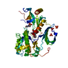
Spodoptera (butterflies/moths) / References: UniProt: P31751

-
Non-polymers , 5 types, 152 molecules

#2: Chemical ChemComp-K06 / 6-[4-(1-azanyl-3-methyl-3-oxidanyl-cyclobutyl)phenyl]-7-phenyl-1-propyl-pyrido[2,3-b][1,4]oxazin-2-one


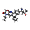
Mass: 443.537 Da / Num. of mol.: 1 / Source method: obtained synthetically / Formula: C27H29N3O3 / Feature type: SUBJECT OF INVESTIGATION
#3: Chemical ChemComp-GOL / GLYCEROL / GLYCERIN / PROPANE-1,2,3-TRIOL


Mass: 92.094 Da / Num. of mol.: 1 / Source method: obtained synthetically / Formula: C3H8O3
#4: Chemical ChemComp-PO4 / PHOSPHATE ION


Mass: 94.971 Da / Num. of mol.: 3 / Source method: obtained synthetically / Formula: PO4
#5: Chemical ChemComp-MES / 2-(N-MORPHOLINO)-ETHANESULFONIC ACID


Mass: 195.237 Da / Num. of mol.: 1 / Source method: obtained synthetically / Formula: C6H13NO4S / Comment: pH buffer*YM
#6: Water ChemComp-HOH / water


Mass: 18.015 Da / Num. of mol.: 146 / Source method: isolated from a natural source / Formula: H2O

-
Details

Has ligand of interestY

-
Experimental details

-
Experiment

ExperimentMethod: X-RAY DIFFRACTION / Number of used crystals: 1

-
Sample preparation

CrystalDensity Matthews: 2.37 Å3/Da / Density % sol: 48 %
Crystal growTemperature: 298 K / Method: vapor diffusion, hanging drop / pH: 6.5 / Details: 0.1 M MES, 1.5 M NaH2PO4

-
Data collection

DiffractionMean temperature: 100 K / Serial crystal experiment: N
Diffraction sourceSource: SYNCHROTRON / Site: SLS / Beamline: X10SA / Wavelength: 1.00004 Å
DetectorType: PSI PILATUS 6M / Detector: PIXEL / Date: Jul 3, 2018
RadiationProtocol: SINGLE WAVELENGTH / Monochromatic (M) / Laue (L): M / Scattering type: x-ray
Radiation wavelengthWavelength: 1.00004 Å / Relative weight: 1
ReflectionResolution: 2.32→61.49 Å / Num. obs: 22371 / % possible obs: 99.58 % / Observed criterion σ(I): 0 / Redundancy: 4.1 % / Biso Wilson estimate: 48.372 Å2 / Rmerge(I) obs: 0.057 / Net I/σ(I): 0
Reflection shellResolution: 2.32→2.57 Å / Redundancy: 4.3 % / Rmerge(I) obs: 0.441 / Num. unique obs: 1613 / % possible all: 99.5

-
Processing

Software
NameVersionClassification
XDSdata reduction
XSCALEdata scaling
REFMAC5.8.0155refinement
MERLOTphasing
RefinementMethod to determine structure: MOLECULAR REPLACEMENT / Resolution: 2.32→61.49 Å / Cor.coef. Fo:Fc: 0.942 / Cor.coef. Fo:Fc free: 0.909 / SU B: 13.585 / SU ML: 0.167 / Cross valid method: THROUGHOUT / ESU R: 0.298 / ESU R Free: 0.24 / Stereochemistry target values: MAXIMUM LIKELIHOOD / Details: HYDROGENS HAVE BEEN ADDED IN THE RIDING POSITIONS
RfactorNum. reflection% reflectionSelection details
Rfree0.2634 1187 5 %RANDOM
Rwork0.20912 ---
obs0.21167 22371 99.58 %-
Solvent computationIon probe radii: 0.8 Å / Shrinkage radii: 0.8 Å / VDW probe radii: 1.2 Å / Solvent model: MASK
Displacement parametersBiso mean: 48.372 Å2
Baniso -1Baniso -2Baniso -3
1-0.12 Å2-0 Å2-0 Å2
2---1.08 Å20 Å2
3---0.96 Å2
Refinement stepCycle: LAST / Resolution: 2.32→61.49 Å
ProteinNucleic acidLigandSolventTotal
Num. atoms3152 0 66 146 3364
Refine LS restraints
Refine-IDTypeDev idealDev ideal targetNumber
X-RAY DIFFRACTIONr_bond_refined_d0.0110.0193237
X-RAY DIFFRACTIONr_bond_other_d0.0040.023110
X-RAY DIFFRACTIONr_angle_refined_deg1.5471.9854368
X-RAY DIFFRACTIONr_angle_other_deg1.21237129
X-RAY DIFFRACTIONr_dihedral_angle_1_deg6.5515380
X-RAY DIFFRACTIONr_dihedral_angle_2_deg30.36622.603146
X-RAY DIFFRACTIONr_dihedral_angle_3_deg13.37415.052573
X-RAY DIFFRACTIONr_dihedral_angle_4_deg20.5411527
X-RAY DIFFRACTIONr_chiral_restr0.0870.2469
X-RAY DIFFRACTIONr_gen_planes_refined0.0050.0213517
X-RAY DIFFRACTIONr_gen_planes_other0.0020.02755
X-RAY DIFFRACTIONr_nbd_refined
X-RAY DIFFRACTIONr_nbd_other
X-RAY DIFFRACTIONr_nbtor_refined
X-RAY DIFFRACTIONr_nbtor_other
X-RAY DIFFRACTIONr_xyhbond_nbd_refined
X-RAY DIFFRACTIONr_xyhbond_nbd_other
X-RAY DIFFRACTIONr_metal_ion_refined
X-RAY DIFFRACTIONr_metal_ion_other
X-RAY DIFFRACTIONr_symmetry_vdw_refined
X-RAY DIFFRACTIONr_symmetry_vdw_other
X-RAY DIFFRACTIONr_symmetry_hbond_refined
X-RAY DIFFRACTIONr_symmetry_hbond_other
X-RAY DIFFRACTIONr_symmetry_metal_ion_refined
X-RAY DIFFRACTIONr_symmetry_metal_ion_other
X-RAY DIFFRACTIONr_mcbond_it2.5723.4731534
X-RAY DIFFRACTIONr_mcbond_other2.5693.4741533
X-RAY DIFFRACTIONr_mcangle_it3.9645.8331909
X-RAY DIFFRACTIONr_mcangle_other3.9655.8331910
X-RAY DIFFRACTIONr_scbond_it3.8064.1531703
X-RAY DIFFRACTIONr_scbond_other3.8024.1531703
X-RAY DIFFRACTIONr_scangle_it
X-RAY DIFFRACTIONr_scangle_other5.6966.7512458
X-RAY DIFFRACTIONr_long_range_B_refined7.91631.3393449
X-RAY DIFFRACTIONr_long_range_B_other7.931.1423428
X-RAY DIFFRACTIONr_rigid_bond_restr
X-RAY DIFFRACTIONr_sphericity_free
X-RAY DIFFRACTIONr_sphericity_bonded
LS refinement shellResolution: 2.32→2.38 Å / Total num. of bins used: 20
RfactorNum. reflection% reflection
Rfree0.404 84 -
Rwork0.316 1613 -
obs--98.84 %
Refinement TLS params.

Method: refined / Refine-ID: X-RAY DIFFRACTION

IDL112)L122)L132)L222)L232)L332)S11 (Å °)S12 (Å °)S13 (Å °)S21 (Å °)S22 (Å °)S23 (Å °)S31 (Å °)S32 (Å °)S33 (Å °)T112)T122)T132)T222)T232)T332)Origin x (Å)Origin y (Å)Origin z (Å)
10.7561-0.1579-0.18425.17193.08744.2387-0.0830.07920.17180.14640.02570.202-0.0807-0.13070.05730.15050.0422-0.01120.1495-0.00230.1321-1.782-13.807-16.746
24.58191.09250.32635.3309-1.28994.8814-0.02860.0331-0.2624-0.0824-0.0217-0.72630.2770.66190.05030.1128-0.0081-0.01230.13990.00410.11191.9831.5457.095
32.1595-0.26570.61252.61170.39064.32990.0371-0.0116-0.04860.0191-0.08360.1181-0.074-0.15890.04640.0896-0.03290.00410.0218-0.00680.0066-12.70615.975-9.6
Refinement TLS group
IDRefine-IDRefine TLS-IDAuth asym-IDAuth seq-ID
1X-RAY DIFFRACTION1A1 - 120
2X-RAY DIFFRACTION2A121 - 228
3X-RAY DIFFRACTION3A229 - 500

+
About Yorodumi

-
News

-
Feb 9, 2022. New format data for meta-information of EMDB entries

New format data for meta-information of EMDB entries

  • Version 3 of the EMDB header file is now the official format.
  • The previous official version 1.9 will be removed from the archive.

Related info.:EMDB header

External links:wwPDB to switch to version 3 of the EMDB data model

-
Aug 12, 2020. Covid-19 info

Covid-19 info

URL: https://pdbjlvh1.pdbj.org/emnavi/covid19.php

New page: Covid-19 featured information page in EM Navigator.

Related info.:Covid-19 info / Mar 5, 2020. Novel coronavirus structure data

+
Mar 5, 2020. Novel coronavirus structure data

Novel coronavirus structure data

Related info.:Yorodumi Speices / Aug 12, 2020. Covid-19 info

External links:COVID-19 featured content - PDBj / Molecule of the Month (242):Coronavirus Proteases

+
Jan 31, 2019. EMDB accession codes are about to change! (news from PDBe EMDB page)

EMDB accession codes are about to change! (news from PDBe EMDB page)

  • The allocation of 4 digits for EMDB accession codes will soon come to an end. Whilst these codes will remain in use, new EMDB accession codes will include an additional digit and will expand incrementally as the available range of codes is exhausted. The current 4-digit format prefixed with “EMD-” (i.e. EMD-XXXX) will advance to a 5-digit format (i.e. EMD-XXXXX), and so on. It is currently estimated that the 4-digit codes will be depleted around Spring 2019, at which point the 5-digit format will come into force.
  • The EM Navigator/Yorodumi systems omit the EMD- prefix.

Related info.:Q: What is EMD? / ID/Accession-code notation in Yorodumi/EM Navigator

External links:EMDB Accession Codes are Changing Soon! / Contact to PDBj

+
Jul 12, 2017. Major update of PDB

Major update of PDB

  • wwPDB released updated PDB data conforming to the new PDBx/mmCIF dictionary.
  • This is a major update changing the version number from 4 to 5, and with Remediation, in which all the entries are updated.
  • In this update, many items about electron microscopy experimental information are reorganized (e.g. em_software).
  • Now, EM Navigator and Yorodumi are based on the updated data.

External links:wwPDB Remediation / Enriched Model Files Conforming to OneDep Data Standards Now Available in the PDB FTP Archive

-
Yorodumi

Thousand views of thousand structures

  • Yorodumi is a browser for structure data from EMDB, PDB, SASBDB, etc.
  • This page is also the successor to EM Navigator detail page, and also detail information page/front-end page for Omokage search.
  • The word "yorodu" (or yorozu) is an old Japanese word meaning "ten thousand". "mi" (miru) is to see.

Related info.:EMDB / PDB / SASBDB / Comparison of 3 databanks / Yorodumi Search / Aug 31, 2016. New EM Navigator & Yorodumi / Yorodumi Papers / Jmol/JSmol / Function and homology information / Changes in new EM Navigator and Yorodumi

Read more