 ムービー
ムービー コントローラー


+ データを開く

- 基本情報
基本情報
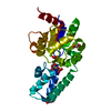
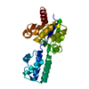
| 登録情報 | データベース: PDB / ID: 8q1c | ||||||||||||
|---|---|---|---|---|---|---|---|---|---|---|---|---|---|
| タイトル | Substrate-free D10N,P146A variant of beta-phosphoglucomutase from Lactococcus lactis | ||||||||||||
 要素 要素 | Beta-phosphoglucomutase | ||||||||||||
 キーワード キーワード | ISOMERASE / mutase | ||||||||||||
| 機能・相同性 |  機能・相同性情報 機能・相同性情報beta-phosphoglucomutase / beta-phosphoglucomutase activity / carbohydrate metabolic process / magnesium ion binding / cytoplasm 類似検索 - 分子機能 | ||||||||||||
| 生物種 |  Lactococcus lactis subsp. lactis Il1403 (乳酸菌) Lactococcus lactis subsp. lactis Il1403 (乳酸菌) | ||||||||||||
| 手法 |  X線回折 / シンクロトロン / 分子置換 / 解像度: 1.679 Å X線回折 / シンクロトロン / 分子置換 / 解像度: 1.679 Å | ||||||||||||
 データ登録者 データ登録者 | Cruz-Navarrete, F.A. / Baxter, N.J. / Flinders, A.J. / Buzoianu, A. / Cliff, M.J. / Baker, P.J. / Waltho, J.P. | ||||||||||||
| 資金援助 |  メキシコ, メキシコ,  英国, 3件 英国, 3件
| ||||||||||||
 引用 引用 | ジャーナル: Commun Biol / 年: 2024 タイトル: Peri active site catalysis of proline isomerisation is the molecular basis of allomorphy in beta-phosphoglucomutase. 著者: Cruz-Navarrete, F.A. / Baxter, N.J. / Flinders, A.J. / Buzoianu, A. / Cliff, M.J. / Baker, P.J. / Waltho, J.P. | ||||||||||||
| 履歴 |
|
- 構造の表示
構造の表示
| 構造ビューア | 分子: MolmilJmol/JSmol |
|---|
- ダウンロードとリンク
-ダウンロード
| PDBx/mmCIF形式 | 8q1c.cif.gz | 106.3 KB | 表示 | PDBx/mmCIF形式 |
|---|---|---|---|---|
| PDB形式 | pdb8q1c.ent.gz | 77.3 KB | 表示 | PDB形式 |
| PDBx/mmJSON形式 | 8q1c.json.gz | ツリー表示 | PDBx/mmJSON形式 | |
| その他 |  その他のダウンロード その他のダウンロード |
-検証レポート
| アーカイブディレクトリ | https://data.pdbj.org/pub/pdb/validation_reports/q1/8q1cftp://data.pdbj.org/pub/pdb/validation_reports/q1/8q1c | HTTPS FTP |
|---|
-関連構造データ

-リンク
 PDBj
PDBj

- 集合体
集合体
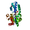
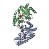
| 登録構造単位 | 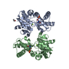
| ||||||||
|---|---|---|---|---|---|---|---|---|---|
| 1 |
| ||||||||
| 単位格子 |
|
-要素
-タンパク質 , 1種, 2分子 AB
| #1: タンパク質 | 分子量: 24212.572 Da / 分子数: 2 / 変異: D10N and P146A / 由来タイプ: 組換発現 由来: (組換発現) Lactococcus lactis subsp. lactis Il1403 (乳酸菌)遺伝子: JCM5805K_2499 / プラスミド: pET22b(+) / 発現宿主: |
|---|
-非ポリマー , 5種, 192分子 


| #2: 化合物 |  分子量: 24.305 Da / 分子数: 2 / 由来タイプ: 合成 / 式: Mg / タイプ: SUBJECT OF INVESTIGATION 分子量: 24.305 Da / 分子数: 2 / 由来タイプ: 合成 / 式: Mg / タイプ: SUBJECT OF INVESTIGATION#3: 化合物 | ChemComp-EDO / |  分子量: 62.068 Da / 分子数: 1 / 由来タイプ: 合成 / 式: C2H6O2 分子量: 62.068 Da / 分子数: 1 / 由来タイプ: 合成 / 式: C2H6O2#4: 化合物 |  分子量: 94.971 Da / 分子数: 2 / 由来タイプ: 合成 / 式: PO4 分子量: 94.971 Da / 分子数: 2 / 由来タイプ: 合成 / 式: PO4#5: 化合物 |  分子量: 122.143 Da / 分子数: 2 / 由来タイプ: 合成 / 式: C4H12NO3 / コメント: pH緩衝剤*YM 分子量: 122.143 Da / 分子数: 2 / 由来タイプ: 合成 / 式: C4H12NO3 / コメント: pH緩衝剤*YM#6: 水 | ChemComp-HOH / | 分子量: 18.015 Da / 分子数: 185 / 由来タイプ: 天然 / 式: H2O |
|---|
-詳細
| 研究の焦点であるリガンドがあるか | Y |
|---|
-実験情報
-実験
| 実験 | 手法: X線回折 / 使用した結晶の数: 1 |
|---|
- 試料調製
試料調製
| 結晶 | マシュー密度: 2.47 Å3/Da / 溶媒含有率: 50.16 % / 解説: rods |
|---|---|
| 結晶化 | 温度: 290 K / 手法: 蒸気拡散法, ハンギングドロップ法 / pH: 7.5 詳細: 0.6 mM bPGM-D10N,P146A 5 mM MgCl2 3 mM AlCl3 20 mM NaF 15 mM glucose 6-phosphate 32 % PEG 4000 200 mM sodium acetate 200 mM tris |
-データ収集
| 回折 | 平均測定温度: 100 K / Serial crystal experiment: N |
|---|---|
| 放射光源 | 由来: シンクロトロン / サイト: Diamond / ビームライン: I03 / 波長: 0.9795 Å |
| 検出器 | タイプ: DECTRIS EIGER2 XE 16M / 検出器: PIXEL / 日付: 2021年6月28日 |
| 放射 | プロトコル: SINGLE WAVELENGTH / 単色(M)・ラウエ(L): M / 散乱光タイプ: x-ray |
| 放射波長 | 波長: 0.9795 Å / 相対比: 1 |
| 反射 | 解像度: 1.679→117.25 Å / Num. obs: 52374 / % possible obs: 98.3 % / 冗長度: 6.5 % / Biso Wilson estimate: 28.2 Å2 / CC1/2: 0.994 / Rmerge(I) obs: 0.089 / Rpim(I) all: 0.04 / Net I/σ(I): 13.8 |
| 反射 シェル | 解像度: 1.68→1.71 Å / 冗長度: 5.7 % / Mean I/σ(I) obs: 0.6 / Num. unique obs: 2284 / CC1/2: 0.277 / % possible all: 86.9 |
- 解析
解析
| ソフトウェア |
| |||||||||||||||||||||||||||||||||||||||||||||||||||||||||||||||||||||||||||||||||||||||||||||||||||||||||||||||||||||||||||||||||||||||||||||||||||||||||||||||||||||||||||||||||||||||||||||||||||||||||||||||||||||||||||||||||||||||
|---|---|---|---|---|---|---|---|---|---|---|---|---|---|---|---|---|---|---|---|---|---|---|---|---|---|---|---|---|---|---|---|---|---|---|---|---|---|---|---|---|---|---|---|---|---|---|---|---|---|---|---|---|---|---|---|---|---|---|---|---|---|---|---|---|---|---|---|---|---|---|---|---|---|---|---|---|---|---|---|---|---|---|---|---|---|---|---|---|---|---|---|---|---|---|---|---|---|---|---|---|---|---|---|---|---|---|---|---|---|---|---|---|---|---|---|---|---|---|---|---|---|---|---|---|---|---|---|---|---|---|---|---|---|---|---|---|---|---|---|---|---|---|---|---|---|---|---|---|---|---|---|---|---|---|---|---|---|---|---|---|---|---|---|---|---|---|---|---|---|---|---|---|---|---|---|---|---|---|---|---|---|---|---|---|---|---|---|---|---|---|---|---|---|---|---|---|---|---|---|---|---|---|---|---|---|---|---|---|---|---|---|---|---|---|---|---|---|---|---|---|---|---|---|---|---|---|---|---|---|---|---|---|
| 精密化 | 構造決定の手法: 分子置換 / 解像度: 1.679→52.702 Å / Cor.coef. Fo:Fc: 0.961 / Cor.coef. Fo:Fc free: 0.917 / WRfactor Rfree: 0.26 / WRfactor Rwork: 0.201 / SU B: 4.146 / SU ML: 0.127 / Average fsc free: 0.9315 / Average fsc work: 0.951 / 交差検証法: FREE R-VALUE / ESU R: 0.116 / ESU R Free: 0.125 / 詳細: Hydrogens have been added in their riding positions
| |||||||||||||||||||||||||||||||||||||||||||||||||||||||||||||||||||||||||||||||||||||||||||||||||||||||||||||||||||||||||||||||||||||||||||||||||||||||||||||||||||||||||||||||||||||||||||||||||||||||||||||||||||||||||||||||||||||||
| 溶媒の処理 | イオンプローブ半径: 0.8 Å / 減衰半径: 0.8 Å / VDWプローブ半径: 1.2 Å / 溶媒モデル: MASK BULK SOLVENT | |||||||||||||||||||||||||||||||||||||||||||||||||||||||||||||||||||||||||||||||||||||||||||||||||||||||||||||||||||||||||||||||||||||||||||||||||||||||||||||||||||||||||||||||||||||||||||||||||||||||||||||||||||||||||||||||||||||||
| 原子変位パラメータ | Biso mean: 39.612 Å2
| |||||||||||||||||||||||||||||||||||||||||||||||||||||||||||||||||||||||||||||||||||||||||||||||||||||||||||||||||||||||||||||||||||||||||||||||||||||||||||||||||||||||||||||||||||||||||||||||||||||||||||||||||||||||||||||||||||||||
| 精密化ステップ | サイクル: LAST / 解像度: 1.679→52.702 Å
| |||||||||||||||||||||||||||||||||||||||||||||||||||||||||||||||||||||||||||||||||||||||||||||||||||||||||||||||||||||||||||||||||||||||||||||||||||||||||||||||||||||||||||||||||||||||||||||||||||||||||||||||||||||||||||||||||||||||
| 拘束条件 |
| |||||||||||||||||||||||||||||||||||||||||||||||||||||||||||||||||||||||||||||||||||||||||||||||||||||||||||||||||||||||||||||||||||||||||||||||||||||||||||||||||||||||||||||||||||||||||||||||||||||||||||||||||||||||||||||||||||||||
| LS精密化 シェル | Refine-ID: X-RAY DIFFRACTION / Total num. of bins used: 20
|
