[English] 日本語
 Yorodumi
- PDB-8puc: Structure of immature HTLV-1 CA-NTD from in vitro assembled MA126... -
+ Open data
- Basic information
| Entry | Database: PDB / ID: 8puc | |||||||||||||||||||||||||||||||||||||||||||||
|---|---|---|---|---|---|---|---|---|---|---|---|---|---|---|---|---|---|---|---|---|---|---|---|---|---|---|---|---|---|---|---|---|---|---|---|---|---|---|---|---|---|---|---|---|---|---|
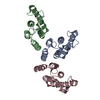
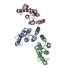
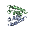
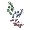
| Title | Structure of immature HTLV-1 CA-NTD from in vitro assembled MA126-CANC tubes: axis angle 05 degrees | |||||||||||||||||||||||||||||||||||||||||||||
 Components Components | Gag protein (Fragment) | |||||||||||||||||||||||||||||||||||||||||||||
 Keywords Keywords | VIRAL PROTEIN / Retrovirus / HTLV / immature capsid / CA / CA-NTD | |||||||||||||||||||||||||||||||||||||||||||||
| Function / homology |  Function and homology information Function and homology information | |||||||||||||||||||||||||||||||||||||||||||||
| Biological species |  Human T-cell leukemia virus type I Human T-cell leukemia virus type I | |||||||||||||||||||||||||||||||||||||||||||||
| Method | ELECTRON MICROSCOPY / subtomogram averaging / cryo EM / Resolution: 4.3 Å | |||||||||||||||||||||||||||||||||||||||||||||
 Authors Authors | Obr, M. / Percipalle, M. / Chernikova, D. / Yang, H. / Thader, A. / Pinke, G. / Porley, D. / Mansky, L.M. / Dick, R.A. / Schur, F.K.M. | |||||||||||||||||||||||||||||||||||||||||||||
| Funding support |  Austria, Austria,  United States, 4items United States, 4items
| |||||||||||||||||||||||||||||||||||||||||||||
 Citation Citation |  Journal: Nat Struct Mol Biol / Year: 2025 Journal: Nat Struct Mol Biol / Year: 2025Title: Distinct stabilization of the human T cell leukemia virus type 1 immature Gag lattice. Authors: Martin Obr / Mathias Percipalle / Darya Chernikova / Huixin Yang / Andreas Thader / Gergely Pinke / Dario Porley / Louis M Mansky / Robert A Dick / Florian K M Schur /    Abstract: Human T cell leukemia virus type 1 (HTLV-1) immature particles differ in morphology from other retroviruses, suggesting a distinct way of assembly. Here we report the results of cryo-electron ...Human T cell leukemia virus type 1 (HTLV-1) immature particles differ in morphology from other retroviruses, suggesting a distinct way of assembly. Here we report the results of cryo-electron tomography studies of HTLV-1 virus-like particles assembled in vitro, as well as derived from cells. This work shows that HTLV-1 uses a distinct mechanism of Gag-Gag interactions to form the immature viral lattice. Analysis of high-resolution structural information from immature capsid (CA) tubular arrays reveals that the primary stabilizing component in HTLV-1 is the N-terminal domain of CA. Mutagenesis analysis supports this observation. This distinguishes HTLV-1 from other retroviruses, in which the stabilization is provided primarily by the C-terminal domain of CA. These results provide structural details of the quaternary arrangement of Gag for an immature deltaretrovirus and this helps explain why HTLV-1 particles are morphologically distinct. | |||||||||||||||||||||||||||||||||||||||||||||
| History |
|
- Structure visualization
| Structure viewer | Molecule:  Molmil Molmil Jmol/JSmol Jmol/JSmol |
|---|
- Downloads & links
- Download
| PDBx/mmCIF format |  8puc.cif.gz 8puc.cif.gz | 53.5 KB | Display |  PDBx/mmCIF format PDBx/mmCIF format |
|---|---|---|---|---|
| PDB format |  pdb8puc.ent.gz pdb8puc.ent.gz | 28.3 KB | Display |  PDB format PDB format |
| PDBx/mmJSON format |  8puc.json.gz 8puc.json.gz | Tree view |  PDBx/mmJSON format PDBx/mmJSON format | |
| Others |  Other downloads Other downloads |
-Validation report
| Summary document |  8puc_validation.pdf.gz 8puc_validation.pdf.gz | 319.1 KB | Display |  wwPDB validaton report wwPDB validaton report |
|---|---|---|---|---|
| Full document |  8puc_full_validation.pdf.gz 8puc_full_validation.pdf.gz | 318.6 KB | Display | |
| Data in XML |  8puc_validation.xml.gz 8puc_validation.xml.gz | 5.6 KB | Display | |
| Data in CIF |  8puc_validation.cif.gz 8puc_validation.cif.gz | 9.1 KB | Display | |
| Arichive directory |  https://data.pdbj.org/pub/pdb/validation_reports/pu/8puc https://data.pdbj.org/pub/pdb/validation_reports/pu/8puc ftp://data.pdbj.org/pub/pdb/validation_reports/pu/8puc ftp://data.pdbj.org/pub/pdb/validation_reports/pu/8puc | HTTPS FTP |
-Related structure data
| Related structure data |  17935MC  8pu6C  8pu7C  8pu8C  8pu9C  8puaC  8pubC  8pudC  8pueC  8pufC  8pugC  8puhC C: citing same article ( M: map data used to model this data |
|---|---|
| Similar structure data | Similarity search - Function & homology  F&H Search F&H Search |
- Links
Links
- Assembly
| Deposited unit | 
|
|---|---|
| 1 |
|
- Components
| #1: Protein | Mass: 13969.809 Da / Num. of mol.: 3 Source method: isolated from a genetically manipulated source Source: (gene. exp.)  Human T-cell leukemia virus type I / Production host: Human T-cell leukemia virus type I / Production host:  Has protein modification | N | |
|---|
-Experimental details
-Experiment
| Experiment | Method: ELECTRON MICROSCOPY |
|---|---|
| EM experiment | Aggregation state: HELICAL ARRAY / 3D reconstruction method: subtomogram averaging |
- Sample preparation
| Component | Name: Human T-cell leukemia virus type I / Type: VIRUS / Entity ID: all / Source: RECOMBINANT | |||||||||||||||||||||||||
|---|---|---|---|---|---|---|---|---|---|---|---|---|---|---|---|---|---|---|---|---|---|---|---|---|---|---|
| Source (natural) | Organism:  Human T-cell leukemia virus type I Human T-cell leukemia virus type I | |||||||||||||||||||||||||
| Source (recombinant) | Organism:  | |||||||||||||||||||||||||
| Details of virus | Empty: YES / Enveloped: NO / Isolate: STRAIN / Type: VIRUS-LIKE PARTICLE | |||||||||||||||||||||||||
| Buffer solution | pH: 8 | |||||||||||||||||||||||||
| Buffer component |
| |||||||||||||||||||||||||
| Specimen | Embedding applied: NO / Shadowing applied: NO / Staining applied: NO / Vitrification applied: YES | |||||||||||||||||||||||||
| Specimen support | Grid material: COPPER / Grid mesh size: 300 divisions/in. / Grid type: C-flat-2/2 | |||||||||||||||||||||||||
| Vitrification | Instrument: LEICA EM GP / Cryogen name: ETHANE / Humidity: 95 % / Chamber temperature: 283 K / Details: Grids coated with 2nm continuous carbon layer |
- Electron microscopy imaging
| Experimental equipment |  Model: Titan Krios / Image courtesy: FEI Company | |||||||||||||||||||||
|---|---|---|---|---|---|---|---|---|---|---|---|---|---|---|---|---|---|---|---|---|---|---|
| EM imaging | Accelerating voltage: 300 kV / Alignment procedure: COMA FREE / Cryogen: NITROGEN / Electron source:
| |||||||||||||||||||||
| Image recording |
| |||||||||||||||||||||
| EM imaging optics |
| |||||||||||||||||||||
| Image scans |
|
- Processing
| EM software |
| ||||||||||||||||||||||||||||||||||||||||||||||||||||||||||||||||||||||||
|---|---|---|---|---|---|---|---|---|---|---|---|---|---|---|---|---|---|---|---|---|---|---|---|---|---|---|---|---|---|---|---|---|---|---|---|---|---|---|---|---|---|---|---|---|---|---|---|---|---|---|---|---|---|---|---|---|---|---|---|---|---|---|---|---|---|---|---|---|---|---|---|---|---|
| Image processing | Details: Datasets (1) and (2) combined during Multiparticle refinement. See Materials and methods for details. | ||||||||||||||||||||||||||||||||||||||||||||||||||||||||||||||||||||||||
| CTF correction | Type: PHASE FLIPPING AND AMPLITUDE CORRECTION | ||||||||||||||||||||||||||||||||||||||||||||||||||||||||||||||||||||||||
| Symmetry | Point symmetry: C2 (2 fold cyclic) | ||||||||||||||||||||||||||||||||||||||||||||||||||||||||||||||||||||||||
| 3D reconstruction | Resolution: 4.3 Å / Resolution method: FSC 0.143 CUT-OFF / Num. of particles: 15800 / Algorithm: BACK PROJECTION / Symmetry type: POINT | ||||||||||||||||||||||||||||||||||||||||||||||||||||||||||||||||||||||||
| EM volume selection | Num. of tomograms: 69 / Num. of volumes extracted: 245000 | ||||||||||||||||||||||||||||||||||||||||||||||||||||||||||||||||||||||||
| Atomic model building | Protocol: RIGID BODY FIT / Space: REAL | ||||||||||||||||||||||||||||||||||||||||||||||||||||||||||||||||||||||||
| Atomic model building | Chain residue range: 13-125 Details: rigid body fit derived from refined model deposited in D_1292131146 Source name: Other / Type: other |
 Movie
Movie Controller


 PDBj
PDBj
 UCSF CHIMERA