 Movie
Movie Controller


+ Open data

- Basic information
| Entry |  | |||||||||||||||
|---|---|---|---|---|---|---|---|---|---|---|---|---|---|---|---|---|
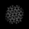
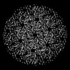
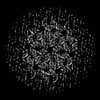
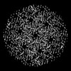
| Title | Cryo-electron tomogram of HTLV-1 MA126CANC tubes | |||||||||||||||
 Map data Map data | cryo-electron tomogram containing HTLV-1 MA126CANC tubes | |||||||||||||||
 Sample Sample |
| |||||||||||||||
 Keywords Keywords | Retrovirus / HTLV / immature capsid / CA / VIRAL PROTEIN | |||||||||||||||
| Biological species |  Human T-cell leukemia virus type I Human T-cell leukemia virus type I | |||||||||||||||
| Method | electron tomography / cryo EM | |||||||||||||||
 Authors Authors | Obr M / Percipalle M / Chernikova D / Yang H / Thader A / Pinke G / Porley D / Mansky LM / Dick RA / Schur FKM | |||||||||||||||
| Funding support |  Austria, Austria,  United States, 4 items United States, 4 items
| |||||||||||||||
 Citation Citation | Journal: Nat Struct Mol Biol / Year: 2025 Title: Distinct stabilization of the human T cell leukemia virus type 1 immature Gag lattice. Authors: Martin Obr / Mathias Percipalle / Darya Chernikova / Huixin Yang / Andreas Thader / Gergely Pinke / Dario Porley / Louis M Mansky / Robert A Dick / Florian K M Schur /  Abstract: Human T cell leukemia virus type 1 (HTLV-1) immature particles differ in morphology from other retroviruses, suggesting a distinct way of assembly. Here we report the results of cryo-electron ...Human T cell leukemia virus type 1 (HTLV-1) immature particles differ in morphology from other retroviruses, suggesting a distinct way of assembly. Here we report the results of cryo-electron tomography studies of HTLV-1 virus-like particles assembled in vitro, as well as derived from cells. This work shows that HTLV-1 uses a distinct mechanism of Gag-Gag interactions to form the immature viral lattice. Analysis of high-resolution structural information from immature capsid (CA) tubular arrays reveals that the primary stabilizing component in HTLV-1 is the N-terminal domain of CA. Mutagenesis analysis supports this observation. This distinguishes HTLV-1 from other retroviruses, in which the stabilization is provided primarily by the C-terminal domain of CA. These results provide structural details of the quaternary arrangement of Gag for an immature deltaretrovirus and this helps explain why HTLV-1 particles are morphologically distinct. | |||||||||||||||
| History |
|
- Structure visualization
| Supplemental images |
|---|

- Downloads & links
-EMDB archive
| Map data | emd_17938.map.gz | 109.5 MB |  EMDB map data format EMDB map data format | |
|---|---|---|---|---|
| Header (meta data) | emd-17938-v30.xmlemd-17938.xml | 18.6 KB 18.6 KB | Display Display | EMDB header |
| Images | 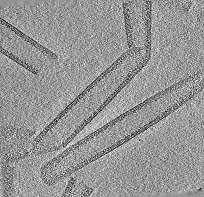 emd_17938.png emd_17938.png | 231.3 KB | ||
| Filedesc metadata | emd-17938.cif.gz | 5.4 KB | ||
| Archive directory |  http://ftp.pdbj.org/pub/emdb/structures/EMD-17938ftp://ftp.pdbj.org/pub/emdb/structures/EMD-17938 http://ftp.pdbj.org/pub/emdb/structures/EMD-17938ftp://ftp.pdbj.org/pub/emdb/structures/EMD-17938 | HTTPS FTP |
-Related structure data
| Related structure data |  8pu6C  8pu7C  8pu8C  8pu9C  8puaC  8pubC  8pucC  8pudC  8pueC  8pufC  8pugC  8puhC C: citing same article ( |
|---|


-Links
| EMDB pages | EMDB (EBI/PDBe) / EMDataResource |
|---|
-Map
| File | Download / File: emd_17938.map.gz / Format: CCP4 / Size: 127.4 MB / Type: IMAGE STORED AS FLOATING POINT NUMBER (4 BYTES) | ||||||||||||||||||||||||||||||||
|---|---|---|---|---|---|---|---|---|---|---|---|---|---|---|---|---|---|---|---|---|---|---|---|---|---|---|---|---|---|---|---|---|---|
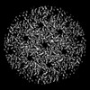
| Annotation | cryo-electron tomogram containing HTLV-1 MA126CANC tubes | ||||||||||||||||||||||||||||||||
| Projections & slices | Image control
Images are generated by Spider. generated in cubic-lattice coordinate | ||||||||||||||||||||||||||||||||
| Voxel size | X=Y=Z: 10.616 Å | ||||||||||||||||||||||||||||||||
| Density |
| ||||||||||||||||||||||||||||||||
| Symmetry | Space group: 1 | ||||||||||||||||||||||||||||||||
| Details | EMDB XML:
|
 Z (Sec.)
 Y (Row.)
 X (Col.)


-Supplemental data
- Sample components
-Entire : Human T-cell leukemia virus type I
| Entire | Name: Human T-cell leukemia virus type I |
|---|---|
| Components |
|
-Supramolecule #1: Human T-cell leukemia virus type I
| Supramolecule | Name: Human T-cell leukemia virus type I / type: virus / ID: 1 / Parent: 0 / Macromolecule list: #1 / NCBI-ID: 11908 / Sci species name: Human T-cell leukemia virus type I / Virus type: VIRUS-LIKE PARTICLE / Virus isolate: STRAIN / Virus enveloped: No / Virus empty: Yes |
|---|
-Experimental details
-Structure determination
| Method | cryo EM |
|---|---|
 Processing Processing | electron tomography |
| Aggregation state | helical array |
-Sample preparation
| Buffer | pH: 8 Component:
| |||||||||||||||
|---|---|---|---|---|---|---|---|---|---|---|---|---|---|---|---|---|
| Grid | Model: C-flat-2/2 / Material: COPPER / Mesh: 300 / Pretreatment - Type: GLOW DISCHARGE / Pretreatment - Time: 30 sec. / Pretreatment - Atmosphere: AMYLAMINE | |||||||||||||||
| Vitrification | Cryogen name: ETHANE / Chamber humidity: 95 % / Chamber temperature: 283 K / Instrument: LEICA EM GP / Details: Grids coated with 2nm continuous carbon layer. | |||||||||||||||
| Sectioning | Other: NO SECTIONING | |||||||||||||||
| Fiducial marker | Manufacturer: Aurion / Diameter: 10 nm |
- Electron microscopy
| Microscope | FEI TITAN KRIOS |
|---|---|
| Specialist optics | Energy filter - Name: GIF Quantum LS / Energy filter - Slit width: 20 eV |
| Image recording | Film or detector model: GATAN K2 QUANTUM (4k x 4k) / Detector mode: COUNTING / Digitization - Dimensions - Width: 3708 pixel / Digitization - Dimensions - Height: 3838 pixel / Average exposure time: 1.05 sec. / Average electron dose: 3.5 e/Å2 |
| Electron beam | Acceleration voltage: 300 kV / Electron source:  FIELD EMISSION GUN FIELD EMISSION GUN |
| Electron optics | Illumination mode: FLOOD BEAM / Imaging mode: BRIGHT FIELD / Cs: 2.7 mm / Nominal defocus max: 3.5 µm / Nominal defocus min: 1.5 µm / Nominal magnification: 105000 |
| Sample stage | Specimen holder model: FEI TITAN KRIOS AUTOGRID HOLDER / Cooling holder cryogen: NITROGEN |
| Experimental equipment |  Model: Titan Krios / Image courtesy: FEI Company |
-Image processing
| Final reconstruction | Algorithm: BACK PROJECTION / Software - Name: IMOD (ver. 4.9.10) / Details: Weighted back projection with SIRT-like filter / Number images used: 41 |
|---|
-Atomic model buiding 1
| Initial model | PDB ID: Chain - Residue range: 13-125 / Chain - Source name: PDB / Chain - Initial model type: experimental model / Details: CA numbering of residues |
|---|---|
| Refinement | Space: REAL / Protocol: AB INITIO MODEL |

