 Movie
Movie Controller


+ Open data

- Basic information
| Entry | Database: PDB / ID: 8pr5 | ||||||||||||
|---|---|---|---|---|---|---|---|---|---|---|---|---|---|
| Title | Structure of the autoinhibited dynactin p150glued projection | ||||||||||||
 Components Components | Dynactin subunit 1 | ||||||||||||
 Keywords Keywords | MOTOR PROTEIN / Dynactin / p150 / LIS1 | ||||||||||||
| Function / homology |  Function and homology information Function and homology informationcentriolar subdistal appendage / centriole-centriole cohesion / positive regulation of neuromuscular junction development / Regulation of PLK1 Activity at G2/M Transition / Loss of Nlp from mitotic centrosomes / Loss of proteins required for interphase microtubule organization from the centrosome / Anchoring of the basal body to the plasma membrane / AURKA Activation by TPX2 / microtubule anchoring at centrosome / Recruitment of mitotic centrosome proteins and complexes ...centriolar subdistal appendage / centriole-centriole cohesion / positive regulation of neuromuscular junction development / Regulation of PLK1 Activity at G2/M Transition / Loss of Nlp from mitotic centrosomes / Loss of proteins required for interphase microtubule organization from the centrosome / Anchoring of the basal body to the plasma membrane / AURKA Activation by TPX2 / microtubule anchoring at centrosome / Recruitment of mitotic centrosome proteins and complexes / nuclear membrane disassembly / ventral spinal cord development / retromer complex / dynein complex / microtubule plus-end / positive regulation of microtubule nucleation / melanosome transport / non-motile cilium assembly / retrograde transport, endosome to Golgi / HSP90 chaperone cycle for steroid hormone receptors (SHR) in the presence of ligand / microtubule associated complex / MHC class II antigen presentation / Recruitment of NuMA to mitotic centrosomes / COPI-mediated anterograde transport / neuromuscular process / nuclear migration / establishment of mitotic spindle orientation / motor behavior / cell leading edge / neuromuscular junction development / intercellular bridge / neuron projection maintenance / centriole / regulation of mitotic spindle organization / neuron cellular homeostasis / kinetochore / spindle pole / mitotic spindle / nuclear envelope / cell cortex / microtubule binding / ciliary basal body / axon / cell division / neuronal cell body / centrosome / protein kinase binding / cytosol Similarity search - Function | ||||||||||||
| Biological species |  | ||||||||||||
| Method | ELECTRON MICROSCOPY / single particle reconstruction / cryo EM / Resolution: 8.6 Å | ||||||||||||
 Authors Authors | Singh, K. / Lau, C.K. / Manigrasso, G. / Gassmann, R. / Carter, A.P. | ||||||||||||
| Funding support |  United Kingdom, European Union, 3items United Kingdom, European Union, 3items
| ||||||||||||
 Citation Citation | Journal: Science / Year: 2024 Title: Molecular mechanism of dynein-dynactin complex assembly by LIS1. Authors: Kashish Singh / Clinton K Lau / Giulia Manigrasso / José B Gama / Reto Gassmann / Andrew P Carter /  Abstract: Cytoplasmic dynein is a microtubule motor vital for cellular organization and division. It functions as a ~4-megadalton complex containing its cofactor dynactin and a cargo-specific coiled-coil ...Cytoplasmic dynein is a microtubule motor vital for cellular organization and division. It functions as a ~4-megadalton complex containing its cofactor dynactin and a cargo-specific coiled-coil adaptor. However, how dynein and dynactin recognize diverse adaptors, how they interact with each other during complex formation, and the role of critical regulators such as lissencephaly-1 (LIS1) protein (LIS1) remain unclear. In this study, we determined the cryo-electron microscopy structure of dynein-dynactin on microtubules with LIS1 and the lysosomal adaptor JIP3. This structure reveals the molecular basis of interactions occurring during dynein activation. We show how JIP3 activates dynein despite its atypical architecture. Unexpectedly, LIS1 binds dynactin's p150 subunit, tethering it along the length of dynein. Our data suggest that LIS1 and p150 constrain dynein-dynactin to ensure efficient complex formation. | ||||||||||||
| History |
|
- Structure visualization
| Structure viewer | Molecule: MolmilJmol/JSmol |
|---|
- Downloads & links
-Download
| PDBx/mmCIF format | 8pr5.cif.gz | 242 KB | Display | PDBx/mmCIF format |
|---|---|---|---|---|
| PDB format | pdb8pr5.ent.gz | Display | PDB format | |
| PDBx/mmJSON format | 8pr5.json.gz | Tree view | PDBx/mmJSON format | |
| Others |  Other downloads Other downloads |
-Validation report
| Arichive directory | https://data.pdbj.org/pub/pdb/validation_reports/pr/8pr5ftp://data.pdbj.org/pub/pdb/validation_reports/pr/8pr5 | HTTPS FTP |
|---|
-Related structure data
| Related structure data | 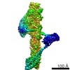 2855M  8pqvC  8pqwC  8pqyC 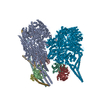 8pqzC  8pr0C  8pr1C  8pr2C  8pr3C  8pr4C  8ptkC M: map data used to model this data C: citing same article ( |
|---|---|
| Similar structure data |


-Links
 PDBj
PDBj- Assembly
| Deposited unit | 
|
|---|---|
| 1 |
|
-Components
| #1: Protein | Mass: 95444.211 Da / Num. of mol.: 2 / Source method: isolated from a natural source / Source: (natural) |
|---|
-Experimental details
-Experiment
| Experiment | Method: ELECTRON MICROSCOPY |
|---|---|
| EM experiment | Aggregation state: PARTICLE / 3D reconstruction method: single particle reconstruction |
- Sample preparation
| Component | Name: dynactin p150 projection / Type: COMPLEX / Entity ID: all / Source: NATURAL |
|---|---|
| Source (natural) | Organism: |
| Buffer solution | pH: 6.5 Details: 50mM KCl, 25mM KH2PO4-K2HPO4, 5mM DDT, 1mM MgCl2, 0.1 mM ATP |
| Specimen | Embedding applied: NO / Shadowing applied: NO / Staining applied: NO / Vitrification applied: YES |
| Vitrification | Cryogen name: NITROGEN |
- Electron microscopy imaging
| Experimental equipment |  Model: Titan Krios / Image courtesy: FEI Company |
|---|---|
| Microscopy | Model: FEI TITAN KRIOS |
| Electron gun | Electron source:  FIELD EMISSION GUN / Accelerating voltage: 300 kV / Illumination mode: FLOOD BEAM FIELD EMISSION GUN / Accelerating voltage: 300 kV / Illumination mode: FLOOD BEAM |
| Electron lens | Mode: BRIGHT FIELD / Nominal defocus max: 7000 nm / Nominal defocus min: 2000 nm |
| Image recording | Electron dose: 51 e/Å2 / Film or detector model: FEI FALCON II (4k x 4k) |
- Processing
| EM software | Name: RELION / Category: final Euler assignment |
|---|---|
| CTF correction | Type: PHASE FLIPPING AND AMPLITUDE CORRECTION |
| 3D reconstruction | Resolution: 8.6 Å / Resolution method: FSC 0.143 CUT-OFF / Num. of particles: 12870 / Symmetry type: POINT |
