 Movie
Movie Controller


+ Open data

- Basic information
| Entry | Database: PDB / ID: 8pqy | ||||||||||||
|---|---|---|---|---|---|---|---|---|---|---|---|---|---|
| Title | Cytoplasmic dynein-1 motor domain bound to LIS1 | ||||||||||||
 Components Components |
| ||||||||||||
 Keywords Keywords | MOTOR PROTEIN / Dynein / AAA-Atpase / p150 / LIS1 | ||||||||||||
| Function / homology |  Function and homology information Function and homology informationmicrotubule cytoskeleton organization involved in establishment of planar polarity / establishment of planar polarity of embryonic epithelium / 1-alkyl-2-acetylglycerophosphocholine esterase complex / corpus callosum morphogenesis / cerebral cortex neuron differentiation / platelet activating factor metabolic process / platelet activating factor catabolic process / establishment of centrosome localization / acrosome assembly / central region of growth cone ...microtubule cytoskeleton organization involved in establishment of planar polarity / establishment of planar polarity of embryonic epithelium / 1-alkyl-2-acetylglycerophosphocholine esterase complex / corpus callosum morphogenesis / cerebral cortex neuron differentiation / platelet activating factor metabolic process / platelet activating factor catabolic process / establishment of centrosome localization / acrosome assembly / central region of growth cone / auditory receptor cell development / microtubule sliding / positive regulation of embryonic development / microtubule organizing center organization / layer formation in cerebral cortex / astral microtubule / cortical microtubule organization / reelin-mediated signaling pathway / positive regulation of dendritic spine morphogenesis / positive regulation of intracellular transport / regulation of metaphase plate congression / positive regulation of spindle assembly / establishment of spindle localization / brain morphogenesis / stereocilium / microtubule plus-end binding / positive regulation of mitotic cell cycle spindle assembly checkpoint / motile cilium / stem cell division / vesicle transport along microtubule / retrograde axonal transport / neuromuscular process controlling balance / COPI-independent Golgi-to-ER retrograde traffic / minus-end-directed microtubule motor activity / microtubule associated complex / P-body assembly / dynein light intermediate chain binding / cytoplasmic dynein complex / kinesin complex / nuclear migration / establishment of mitotic spindle orientation / dynein intermediate chain binding / neuroblast proliferation / germ cell development / cell leading edge / transmission of nerve impulse / dynein complex binding / dynactin binding / cochlea development / positive regulation of axon extension / microtubule-based process / COPI-mediated anterograde transport / cytoplasmic microtubule / positive regulation of mitotic cell cycle / phospholipase binding / adult locomotory behavior / cytoplasmic microtubule organization / axon cytoplasm / Loss of Nlp from mitotic centrosomes / Loss of proteins required for interphase microtubule organization from the centrosome / Amplification of signal from unattached kinetochores via a MAD2 inhibitory signal / Recruitment of mitotic centrosome proteins and complexes / MHC class II antigen presentation / Recruitment of NuMA to mitotic centrosomes / Anchoring of the basal body to the plasma membrane / HSP90 chaperone cycle for steroid hormone receptors (SHR) in the presence of ligand / Mitotic Prometaphase / EML4 and NUDC in mitotic spindle formation / AURKA Activation by TPX2 / male germ cell nucleus / Resolution of Sister Chromatid Cohesion / stress granule assembly / mitotic spindle organization / regulation of mitotic spindle organization / hippocampus development / regulation of microtubule cytoskeleton organization / filopodium / phosphoprotein binding / RHO GTPases Activate Formins / cerebral cortex development / microtubule cytoskeleton organization / modulation of chemical synaptic transmission / neuron migration / kinetochore / Schaffer collateral - CA1 synapse / HCMV Early Events / Aggrephagy / azurophil granule lumen / Separation of Sister Chromatids / Regulation of PLK1 Activity at G2/M Transition / nuclear envelope / negative regulation of neuron projection development / heparin binding / positive regulation of cold-induced thermogenesis / actin cytoskeleton organization / nuclear membrane / cell cortex / chemical synaptic transmission / microtubule binding / microtubule Similarity search - Function | ||||||||||||
| Biological species |  Homo sapiens (human) Homo sapiens (human) | ||||||||||||
| Method | ELECTRON MICROSCOPY / single particle reconstruction / cryo EM / Resolution: 3.8 Å | ||||||||||||
 Authors Authors | Singh, K. / Lau, C.K. / Manigrasso, G. / Gassmann, R. / Carter, A.P. | ||||||||||||
| Funding support |  United Kingdom, European Union, 3items United Kingdom, European Union, 3items
| ||||||||||||
 Citation Citation | Journal: Science / Year: 2024 Title: Molecular mechanism of dynein-dynactin complex assembly by LIS1. Authors: Kashish Singh / Clinton K Lau / Giulia Manigrasso / José B Gama / Reto Gassmann / Andrew P Carter /  Abstract: Cytoplasmic dynein is a microtubule motor vital for cellular organization and division. It functions as a ~4-megadalton complex containing its cofactor dynactin and a cargo-specific coiled-coil ...Cytoplasmic dynein is a microtubule motor vital for cellular organization and division. It functions as a ~4-megadalton complex containing its cofactor dynactin and a cargo-specific coiled-coil adaptor. However, how dynein and dynactin recognize diverse adaptors, how they interact with each other during complex formation, and the role of critical regulators such as lissencephaly-1 (LIS1) protein (LIS1) remain unclear. In this study, we determined the cryo-electron microscopy structure of dynein-dynactin on microtubules with LIS1 and the lysosomal adaptor JIP3. This structure reveals the molecular basis of interactions occurring during dynein activation. We show how JIP3 activates dynein despite its atypical architecture. Unexpectedly, LIS1 binds dynactin's p150 subunit, tethering it along the length of dynein. Our data suggest that LIS1 and p150 constrain dynein-dynactin to ensure efficient complex formation. | ||||||||||||
| History |
|
- Structure visualization
| Structure viewer | Molecule: MolmilJmol/JSmol |
|---|
- Downloads & links
-Download
| PDBx/mmCIF format | 8pqy.cif.gz | 662.7 KB | Display | PDBx/mmCIF format |
|---|---|---|---|---|
| PDB format | pdb8pqy.ent.gz | Display | PDB format | |
| PDBx/mmJSON format | 8pqy.json.gz | Tree view | PDBx/mmJSON format | |
| Others |  Other downloads Other downloads |
-Validation report
| Arichive directory | https://data.pdbj.org/pub/pdb/validation_reports/pq/8pqyftp://data.pdbj.org/pub/pdb/validation_reports/pq/8pqy | HTTPS FTP |
|---|
-Related structure data
| Related structure data |  17828MC  8pqvC  8pqwC 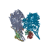 8pqzC  8pr0C  8pr1C  8pr2C  8pr3C  8pr4C  8pr5C  8ptkC M: map data used to model this data C: citing same article ( |
|---|---|
| Similar structure data |


-Links
 PDBj
PDBj


- Assembly
| Deposited unit | 
|
|---|---|
| 1 |
|
-Components
| #1: Protein | Mass: 533055.125 Da / Num. of mol.: 1 / Mutation: R1567E, K1610E Source method: isolated from a genetically manipulated source Source: (gene. exp.) Homo sapiens (human) / Gene: DYNC1H1, DHC1, DNCH1, DNCL, DNECL, DYHC, KIAA0325 / Production host:   Spodoptera frugiperda (fall armyworm) / References: UniProt: Q14204 Spodoptera frugiperda (fall armyworm) / References: UniProt: Q14204 | ||||||||
|---|---|---|---|---|---|---|---|---|---|
| #2: Protein | Mass: 46709.984 Da / Num. of mol.: 2 Source method: isolated from a genetically manipulated source Source: (gene. exp.) Homo sapiens (human) / Gene: PAFAH1B1, LIS1, MDCR, MDS, PAFAHA / Production host: Spodoptera frugiperda (fall armyworm) / References: UniProt: P43034#3: Chemical |   Mass: 427.201 Da / Num. of mol.: 3 / Source method: obtained synthetically / Formula: C10H15N5O10P2 / Feature type: SUBJECT OF INVESTIGATION / Comment: ADP, energy-carrying molecule*YM Mass: 427.201 Da / Num. of mol.: 3 / Source method: obtained synthetically / Formula: C10H15N5O10P2 / Feature type: SUBJECT OF INVESTIGATION / Comment: ADP, energy-carrying molecule*YM#4: Chemical |   Mass: 24.305 Da / Num. of mol.: 2 / Source method: obtained synthetically / Formula: Mg Mass: 24.305 Da / Num. of mol.: 2 / Source method: obtained synthetically / Formula: Mg#5: Chemical | ChemComp-ATP / |   Mass: 507.181 Da / Num. of mol.: 1 / Source method: obtained synthetically / Formula: C10H16N5O13P3 / Feature type: SUBJECT OF INVESTIGATION / Comment: ATP, energy-carrying molecule*YM Mass: 507.181 Da / Num. of mol.: 1 / Source method: obtained synthetically / Formula: C10H16N5O13P3 / Feature type: SUBJECT OF INVESTIGATION / Comment: ATP, energy-carrying molecule*YMHas ligand of interest | Y | |
-Experimental details
-Experiment
| Experiment | Method: ELECTRON MICROSCOPY |
|---|---|
| EM experiment | Aggregation state: PARTICLE / 3D reconstruction method: single particle reconstruction |
- Sample preparation
| Component | Name: Cytoplasmic dynein-1 bound to LIS1 / Type: COMPLEX / Entity ID: #1-#2 / Source: RECOMBINANT |
|---|---|
| Source (natural) | Organism: Homo sapiens (human) |
| Source (recombinant) | Organism: Spodoptera frugiperda (fall armyworm) |
| Buffer solution | pH: 7.2 |
| Specimen | Embedding applied: NO / Shadowing applied: NO / Staining applied: NO / Vitrification applied: YES |
| Vitrification | Cryogen name: ETHANE |
- Electron microscopy imaging
| Experimental equipment |  Model: Titan Krios / Image courtesy: FEI Company |
|---|---|
| Microscopy | Model: FEI TITAN KRIOS |
| Electron gun | Electron source:  FIELD EMISSION GUN / Accelerating voltage: 300 kV / Illumination mode: FLOOD BEAM FIELD EMISSION GUN / Accelerating voltage: 300 kV / Illumination mode: FLOOD BEAM |
| Electron lens | Mode: BRIGHT FIELD / Nominal defocus max: 4000 nm / Nominal defocus min: 500 nm |
| Image recording | Electron dose: 53 e/Å2 / Film or detector model: GATAN K3 BIOQUANTUM (6k x 4k) |
- Processing
| EM software | Name: PHENIX / Version: 1.20_4459: / Category: model refinement | ||||||||||||||||||||||||
|---|---|---|---|---|---|---|---|---|---|---|---|---|---|---|---|---|---|---|---|---|---|---|---|---|---|
| CTF correction | Type: PHASE FLIPPING AND AMPLITUDE CORRECTION | ||||||||||||||||||||||||
| 3D reconstruction | Resolution: 3.8 Å / Resolution method: FSC 0.143 CUT-OFF / Num. of particles: 90594 / Symmetry type: POINT | ||||||||||||||||||||||||
| Atomic model building | PDB-ID: 7Z8G Accession code: 7Z8G / Source name: PDB / Type: experimental model | ||||||||||||||||||||||||
| Refine LS restraints |
|

