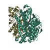
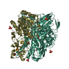
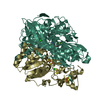
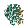
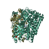
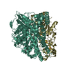
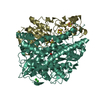
Entry Database : PDB / ID : 8pouTitle Crystal Structure of the C19G/C120G variant of the membrane-bound [NiFe]-Hydrogenase from Cupriavidus necator in the air-oxidized state at 1.65 A Resolution. (Uptake hydrogenase ...) x 2 Keywords / / / / / / / / / / / / / / / / / / / / / / / Function / homology Function Domain/homology Component
/ / / / / / / / / / / / / / / / / / / / / / / / / / / / / / / / / / / / / / / / / / / / / / / / / Biological species Cupriavidus necator H16 (bacteria)Method / / / Resolution : 1.65 Å Authors Schmidt, A. / Kalms, J. / Scheerer, P. Funding support Organization Grant number Country German Research Foundation (DFG) DFG Cluster of Excellence Unifying Concepts in Catalysis - Research Field D3/E3-1 German Research Foundation (DFG) UniSysCat - Germany Excellence Strategy EXC2008/1-390540038
Journal : Chem Sci / Year : 2023Title : Stepwise conversion of the Cys 6 [4Fe-3S] to a Cys 4 [4Fe-4S] cluster and its impact on the oxygen tolerance of [NiFe]-hydrogenase.Authors : Schmidt, A. / Kalms, J. / Lorent, C. / Katz, S. / Frielingsdorf, S. / Evans, R.M. / Fritsch, J. / Siebert, E. / Teutloff, C. / Armstrong, F.A. / Zebger, I. / Lenz, O. / Scheerer, P. History Deposition Jul 5, 2023 Deposition site / Processing site Revision 1.0 Nov 1, 2023 Provider / Type Revision 1.1 Feb 7, 2024 Group / Category / Item
Show all Show less
 Movie
Movie Controller

 Yorodumi
 Open data
 Basic information
 Components
 Keywords
 Function and homology information
 Cupriavidus necator H16 (bacteria)
 X-RAY DIFFRACTION /
  Authors
 Germany, 2items
  Citation
 Structure visualization
 Downloads & links
 Other downloads
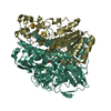


 PDBj
PDBj


 Assembly


 Mass: 210.583 Da / Num. of mol.: 1 / Source method: obtained synthetically / Formula: C3FeN2NiO2 / Feature type: SUBJECT OF INVESTIGATION
 Mass: 24.305 Da / Num. of mol.: 1 / Source method: obtained synthetically / Formula: Mg
 Mass: 35.453 Da / Num. of mol.: 3 / Source method: obtained synthetically / Formula: Cl
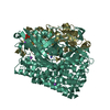
 Mass: 351.640 Da / Num. of mol.: 2 / Source method: obtained synthetically / Formula: Fe4S4 / Feature type: SUBJECT OF INVESTIGATION
 Mass: 295.795 Da / Num. of mol.: 1 / Source method: obtained synthetically / Formula: Fe3S4
 Sample preparation
 / Beamline: ID29 / Wavelength: 0.97625 Å
 Processing