 Movie
Movie Controller

[English] 日本語
 Yorodumi
- PDB-8pf0: Diubiquitin-derived artificial binding protein (Affilin) variant ... -
+ Open data

- Basic information
| Entry | Database: PDB / ID: 8pf0 | ||||||
|---|---|---|---|---|---|---|---|
| Title | Diubiquitin-derived artificial binding protein (Affilin) variant Af1 specific to oncofetal fibronectin | ||||||
 Components Components | Affilin variant Af1 (77405) | ||||||
 Keywords Keywords | DE NOVO PROTEIN / extra domain B / EDB / oncofetal fibronectin / ubiquitin / diubiqutin / Affilin / beta strand register shift / strand slippage / scaffold / artificial binding protein / plasticity / directed evolution | ||||||
| Function / homology | COPPER (II) ION / IMIDAZOLE Function and homology information Function and homology information | ||||||
| Biological species |  Homo sapiens (human) Homo sapiens (human) | ||||||
| Method |  X-RAY DIFFRACTION / SYNCHROTRON / SAD / Resolution: 2.2 Å X-RAY DIFFRACTION / SYNCHROTRON / SAD / Resolution: 2.2 Å | ||||||
 Authors Authors | Parthier, C. / Katzschmann, A. / Fiedler, E. / Haupts, U. / Reimann, A. | ||||||
| Funding support | 1items
| ||||||
 Citation Citation | Journal: Commun Biol / Year: 2024 Title: Ubiquitin-derived artificial binding proteins targeting oncofetal fibronectin reveal scaffold plasticity by beta-strand slippage. Authors: Katzschmann, A. / Haupts, U. / Reimann, A. / Settele, F. / Gloser-Braunig, M. / Fiedler, E. / Parthier, C. | ||||||
| History |
|
- Structure visualization
| Structure viewer | Molecule: MolmilJmol/JSmol |
|---|
- Downloads & links
-Download
| PDBx/mmCIF format | 8pf0.cif.gz | 80.4 KB | Display | PDBx/mmCIF format |
|---|---|---|---|---|
| PDB format | pdb8pf0.ent.gz | 59 KB | Display | PDB format |
| PDBx/mmJSON format | 8pf0.json.gz | Tree view | PDBx/mmJSON format | |
| Others |  Other downloads Other downloads |
-Validation report
| Summary document | 8pf0_validation.pdf.gz | 437.1 KB | Display | wwPDB validaton report |
|---|---|---|---|---|
| Full document | 8pf0_full_validation.pdf.gz | 436.9 KB | Display | |
| Data in XML | 8pf0_validation.xml.gz | 8.7 KB | Display | |
| Data in CIF | 8pf0_validation.cif.gz | 11.2 KB | Display | |
| Arichive directory | https://data.pdbj.org/pub/pdb/validation_reports/pf/8pf0ftp://data.pdbj.org/pub/pdb/validation_reports/pf/8pf0 | HTTPS FTP |
-Related structure data

-Links
 PDBj
PDBj


- Assembly
| Deposited unit | 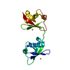
| ||||||||
|---|---|---|---|---|---|---|---|---|---|
| 1 |
| ||||||||
| Unit cell |
|
-Components
| #1: Protein | Mass: 17529.107 Da / Num. of mol.: 1 Source method: isolated from a genetically manipulated source Source: (gene. exp.) Homo sapiens (human) / Production host:  | ||||||
|---|---|---|---|---|---|---|---|
| #2: Chemical | ChemComp-CU /   Mass: 63.546 Da / Num. of mol.: 4 Mass: 63.546 Da / Num. of mol.: 4Source method: isolated from a genetically manipulated source Formula: Cu #3: Chemical |   Mass: 69.085 Da / Num. of mol.: 2 / Source method: obtained synthetically / Formula: C3H5N2 Mass: 69.085 Da / Num. of mol.: 2 / Source method: obtained synthetically / Formula: C3H5N2#4: Water | ChemComp-HOH / |  Mass: 18.015 Da / Num. of mol.: 78 / Source method: isolated from a natural source / Formula: H2O Mass: 18.015 Da / Num. of mol.: 78 / Source method: isolated from a natural source / Formula: H2OHas ligand of interest | N | |
-Experimental details
-Experiment
| Experiment | Method: X-RAY DIFFRACTION / Number of used crystals: 1 |
|---|
- Sample preparation
| Crystal | Density Matthews: 3.81 Å3/Da / Density % sol: 67.73 % |
|---|---|
| Crystal grow | Temperature: 288 K / Method: vapor diffusion, hanging drop / pH: 6 Details: 100 mM Imidazol/MES, 60 mM calcium/magnesium chloride, 30% PEG 8000/ethylene glycol (w/v), 10 mM copper(II)chloride dehydrate |
-Data collection
| Diffraction | Mean temperature: 100 K / Serial crystal experiment: N | ||||||||||||||||||||||||||||||||||||||||||||||||||||||||||||||||||||||||
|---|---|---|---|---|---|---|---|---|---|---|---|---|---|---|---|---|---|---|---|---|---|---|---|---|---|---|---|---|---|---|---|---|---|---|---|---|---|---|---|---|---|---|---|---|---|---|---|---|---|---|---|---|---|---|---|---|---|---|---|---|---|---|---|---|---|---|---|---|---|---|---|---|---|
| Diffraction source | Source: SYNCHROTRON / Site: BESSY  / Beamline: 14.2 / Wavelength: 0.9184 Å / Beamline: 14.2 / Wavelength: 0.9184 Å | ||||||||||||||||||||||||||||||||||||||||||||||||||||||||||||||||||||||||
| Detector | Type: MARMOSAIC 225 mm CCD / Detector: CCD / Date: May 28, 2013 | ||||||||||||||||||||||||||||||||||||||||||||||||||||||||||||||||||||||||
| Radiation | Protocol: SINGLE WAVELENGTH / Monochromatic (M) / Laue (L): M / Scattering type: x-ray | ||||||||||||||||||||||||||||||||||||||||||||||||||||||||||||||||||||||||
| Radiation wavelength | Wavelength: 0.9184 Å / Relative weight: 1 | ||||||||||||||||||||||||||||||||||||||||||||||||||||||||||||||||||||||||
| Reflection | Resolution: 2.2→20 Å / Num. obs: 26176 / % possible obs: 99.5 % / Redundancy: 3.8 % / CC1/2: 0.999 / Rmerge(I) obs: 0.031 / Rrim(I) all: 0.036 / Net I/σ(I): 22.88 | ||||||||||||||||||||||||||||||||||||||||||||||||||||||||||||||||||||||||
| Reflection shell |
|
- Processing
| Software |
| ||||||||||||||||||||||||||||||||||||||||||||||||||||||||||||||||||||||||||||||||||||||||||||||||||||
|---|---|---|---|---|---|---|---|---|---|---|---|---|---|---|---|---|---|---|---|---|---|---|---|---|---|---|---|---|---|---|---|---|---|---|---|---|---|---|---|---|---|---|---|---|---|---|---|---|---|---|---|---|---|---|---|---|---|---|---|---|---|---|---|---|---|---|---|---|---|---|---|---|---|---|---|---|---|---|---|---|---|---|---|---|---|---|---|---|---|---|---|---|---|---|---|---|---|---|---|---|---|
| Refinement | Method to determine structure: SAD / Resolution: 2.2→19.05 Å / SU ML: 0.36 / Cross valid method: FREE R-VALUE / σ(F): 1.17 / Phase error: 29.9 / Stereochemistry target values: ML
| ||||||||||||||||||||||||||||||||||||||||||||||||||||||||||||||||||||||||||||||||||||||||||||||||||||
| Solvent computation | Shrinkage radii: 0.9 Å / VDW probe radii: 1.11 Å / Solvent model: FLAT BULK SOLVENT MODEL | ||||||||||||||||||||||||||||||||||||||||||||||||||||||||||||||||||||||||||||||||||||||||||||||||||||
| Refinement step | Cycle: LAST / Resolution: 2.2→19.05 Å
| ||||||||||||||||||||||||||||||||||||||||||||||||||||||||||||||||||||||||||||||||||||||||||||||||||||
| Refine LS restraints |
| ||||||||||||||||||||||||||||||||||||||||||||||||||||||||||||||||||||||||||||||||||||||||||||||||||||
| LS refinement shell |
| ||||||||||||||||||||||||||||||||||||||||||||||||||||||||||||||||||||||||||||||||||||||||||||||||||||
| Refinement TLS params. | Method: refined / Refine-ID: X-RAY DIFFRACTION
| ||||||||||||||||||||||||||||||||||||||||||||||||||||||||||||||||||||||||||||||||||||||||||||||||||||
| Refinement TLS group |
|
