 Movie
Movie Controller

[English] 日本語
 Yorodumi
- PDB-8pdk: X-ray structure of the Thermus thermophilus PilF-GSPIIB domain in... -
+ Open data

- Basic information
| Entry | Database: PDB / ID: 8pdk | ||||||
|---|---|---|---|---|---|---|---|
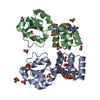
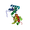
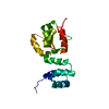
| Title | X-ray structure of the Thermus thermophilus PilF-GSPIIB domain in the c-di-GMP bound state | ||||||
 Components Components | c-di-GMP binding domain of the ATPase enzyme PilF | ||||||
 Keywords Keywords | MOTOR PROTEIN / PilT class / GSPII / ligand binding / c-di-GMP | ||||||
| Function / homology |  Function and homology information Function and homology information | ||||||
| Biological species |   Thermus thermophilus HB27 (bacteria) Thermus thermophilus HB27 (bacteria) | ||||||
| Method |  X-RAY DIFFRACTION / SYNCHROTRON / MOLECULAR REPLACEMENT / Resolution: 2 Å X-RAY DIFFRACTION / SYNCHROTRON / MOLECULAR REPLACEMENT / Resolution: 2 Å | ||||||
 Authors Authors | Neissner, K. / Woehnert, J. | ||||||
| Funding support |  Germany, 1items Germany, 1items
| ||||||
 Citation Citation | Journal: J.Biol.Chem. / Year: 2024 Title: The structural basis for high-affinity c-di-GMP binding to the GSPII-B domain of the traffic ATPase PilF from Thermus thermophilus. Authors: Neissner, K. / Keller, H. / Kirchner, L. / Dusterhus, S. / Duchardt-Ferner, E. / Averhoff, B. / Wohnert, J. | ||||||
| History |
|
- Structure visualization
| Structure viewer | Molecule: MolmilJmol/JSmol |
|---|
- Downloads & links
-Download
| PDBx/mmCIF format | 8pdk.cif.gz | 162.6 KB | Display | PDBx/mmCIF format |
|---|---|---|---|---|
| PDB format | pdb8pdk.ent.gz | 106.5 KB | Display | PDB format |
| PDBx/mmJSON format | 8pdk.json.gz | Tree view | PDBx/mmJSON format | |
| Others |  Other downloads Other downloads |
-Validation report
| Arichive directory | https://data.pdbj.org/pub/pdb/validation_reports/pd/8pdkftp://data.pdbj.org/pub/pdb/validation_reports/pd/8pdk | HTTPS FTP |
|---|
-Related structure data

-Links
 PDBj
PDBj

- Assembly
| Deposited unit | 
| ||||||||||
|---|---|---|---|---|---|---|---|---|---|---|---|
| 1 |
| ||||||||||
| Unit cell |
| ||||||||||
| Components on special symmetry positions |
|
-Components
| #1: Protein | Mass: 16269.562 Da / Num. of mol.: 2 Source method: isolated from a genetically manipulated source Source: (gene. exp.) Thermus thermophilus HB27 (bacteria) / Gene: pilF, TT_C1622 / Production host: #2: Chemical |   Mass: 690.411 Da / Num. of mol.: 2 / Source method: obtained synthetically / Formula: C20H24N10O14P2 / Feature type: SUBJECT OF INVESTIGATION Mass: 690.411 Da / Num. of mol.: 2 / Source method: obtained synthetically / Formula: C20H24N10O14P2 / Feature type: SUBJECT OF INVESTIGATION#3: Chemical | ChemComp-SO4 /   Mass: 96.063 Da / Num. of mol.: 7 / Source method: obtained synthetically / Formula: SO4 Mass: 96.063 Da / Num. of mol.: 7 / Source method: obtained synthetically / Formula: SO4#4: Chemical | ChemComp-ACT /   Mass: 59.044 Da / Num. of mol.: 13 / Source method: obtained synthetically / Formula: C2H3O2 Mass: 59.044 Da / Num. of mol.: 13 / Source method: obtained synthetically / Formula: C2H3O2#5: Water | ChemComp-HOH / |  Mass: 18.015 Da / Num. of mol.: 178 / Source method: isolated from a natural source / Formula: H2O Mass: 18.015 Da / Num. of mol.: 178 / Source method: isolated from a natural source / Formula: H2OHas ligand of interest | Y | Has protein modification | N | |
|---|
-Experimental details
-Experiment
| Experiment | Method: X-RAY DIFFRACTION / Number of used crystals: 1 |
|---|
- Sample preparation
| Crystal | Density Matthews: 2.92 Å3/Da / Density % sol: 57.87 % |
|---|---|
| Crystal grow | Temperature: 291 K / Method: vapor diffusion, hanging drop / pH: 5.5 / Details: 1.8 M Lithiumsulfate 0.1 M Sodiumacetate pH 5.5 |
-Data collection
| Diffraction | Mean temperature: 100 K / Serial crystal experiment: N |
|---|---|
| Diffraction source | Source: SYNCHROTRON / Site: NSLS  / Beamline: X6A / Wavelength: 1 Å / Beamline: X6A / Wavelength: 1 Å |
| Detector | Type: DECTRIS PILATUS 2M-F / Detector: PIXEL / Date: Dec 14, 2019 |
| Radiation | Protocol: MAD / Monochromatic (M) / Laue (L): M / Scattering type: x-ray |
| Radiation wavelength | Wavelength: 1 Å / Relative weight: 1 |
| Reflection | Resolution: 2→41.33 Å / Num. obs: 25934 / % possible obs: 99.96 % / Redundancy: 5.2 % / Biso Wilson estimate: 37.88 Å2 / CC1/2: 1 / Rmerge(I) obs: 0.03878 / Rpim(I) all: 0.01886 / Rrim(I) all: 0.04317 / Net I/σ(I): 29.09 |
| Reflection shell | Resolution: 2→2.071 Å / Rmerge(I) obs: 0.6646 / Mean I/σ(I) obs: 2.6 / Num. unique obs: 2624 / CC1/2: 0.779 / Rpim(I) all: 0.3309 / Rrim(I) all: 0.7432 / % possible all: 99.96 |
- Processing
| Software |
| ||||||||||||||||||||||||||||||||||||||||||||||||||||||||||||||||||||||||||||||||||||
|---|---|---|---|---|---|---|---|---|---|---|---|---|---|---|---|---|---|---|---|---|---|---|---|---|---|---|---|---|---|---|---|---|---|---|---|---|---|---|---|---|---|---|---|---|---|---|---|---|---|---|---|---|---|---|---|---|---|---|---|---|---|---|---|---|---|---|---|---|---|---|---|---|---|---|---|---|---|---|---|---|---|---|---|---|---|
| Refinement | Method to determine structure: MOLECULAR REPLACEMENT / Resolution: 2→41.33 Å / SU ML: 0.2417 / Cross valid method: FREE R-VALUE / σ(F): 1.97 / Phase error: 23.4766 Stereochemistry target values: GeoStd + Monomer Library + CDL v1.2
| ||||||||||||||||||||||||||||||||||||||||||||||||||||||||||||||||||||||||||||||||||||
| Solvent computation | Shrinkage radii: 0.9 Å / VDW probe radii: 1.11 Å / Solvent model: FLAT BULK SOLVENT MODEL | ||||||||||||||||||||||||||||||||||||||||||||||||||||||||||||||||||||||||||||||||||||
| Displacement parameters | Biso mean: 46.62 Å2 | ||||||||||||||||||||||||||||||||||||||||||||||||||||||||||||||||||||||||||||||||||||
| Refinement step | Cycle: LAST / Resolution: 2→41.33 Å
| ||||||||||||||||||||||||||||||||||||||||||||||||||||||||||||||||||||||||||||||||||||
| Refine LS restraints |
| ||||||||||||||||||||||||||||||||||||||||||||||||||||||||||||||||||||||||||||||||||||
| LS refinement shell |
| ||||||||||||||||||||||||||||||||||||||||||||||||||||||||||||||||||||||||||||||||||||
| Refinement TLS params. | Method: refined / Origin x: -22.2051116464 Å / Origin y: 22.9772054972 Å / Origin z: 6.8706395989 Å
| ||||||||||||||||||||||||||||||||||||||||||||||||||||||||||||||||||||||||||||||||||||
| Refinement TLS group | Selection details: all |
