Type II secretion system protein GspE, N-terminal / MshEN domain / Type II secretion system protein GspE, N-terminal superfamily / Bacterial type II secretion system protein E signature. / Type II/IV secretion system protein / Type II/IV secretion system protein / P-loop containing nucleoside triphosphate hydrolase Similarity search - Domain/homology
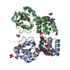
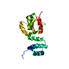
ATP-bindingmotif-containingproteinpilF / c-di-GMP binding domain of the ATPase enzyme PilF
Mass: 16269.562 Da / Num. of mol.: 1 Source method: isolated from a genetically manipulated source Details: Each molecule consists of the protein bound to c-di-GMP (residues 147-148). Source: (gene. exp.) Thermus thermophilus HB27 (bacteria) / Gene: pilF, TT_C1622 / Production host: Escherichia coli BL21(DE3) (bacteria) / References: UniProt: Q72H73
leucine and valine methylgroups (CD1/CD2) (CG1/CG2) were stereospecifically 13C labelled. For expression a mixture of 10%-13C-labelled and 90%-unlabelled glucose was used to achieve stereospecific labelling.
solution
5
545 uM [U-100% 15N] PilF159-302, 700 uM c-di-GMP, 90% H2O/10% D2O
15N_PilF159-302_unlabelled-c-di-GMP
90% H2O/10% D2O
solution
6
420 uM PilF159-302, 420 uM [U-13C; U-15N] c-di-GMP, 90% H2O/10% D2O
PilF159-302_13C_15N-c-di-GMP
90% H2O/10% D2O
solution
7
940 uM [U-100% 15N] PilF159-302, 1122 uM [U-13C; U-15N] c-di-GMP, 90% H2O/10% D2O
15N_PilF159-302_13C_15N-c-di-GMP
90% H2O/10% D2O
Sample
Conc. (mg/ml)
Component
Isotopic labeling
Solution-ID
541uM
PilF159-302
[U-13C; U-15N]
1
812uM
c-di-GMP
naturalabundance
1
460ug/uL
PilF159-302
[U-100% 15N]
2
707uM
c-di-GMP
naturalabundance
2
545uM
PilF159-302
[U-15N]-Leu/Val-13C
3
818uM
c-di-GMP
naturalabundance
3
522uM
PilF159-302
[U-15N]-Leu/Val-13C-stereospecific
4
627uM
c-di-GMP
naturalabundance
4
545uM
PilF159-302
[U-100% 15N]
5
700uM
c-di-GMP
naturalabundance
5
420uM
PilF159-302
naturalabundance
6
420uM
c-di-GMP
[U-13C; U-15N]
6
940uM
PilF159-302
[U-100% 15N]
7
1122uM
c-di-GMP
[U-13C; U-15N]
7
Sample conditions
Ionic strength: 200 mM NaCl mM / Label: conditions_1 / pH: 5.8 / Pressure: ambient atm / Temperature: 318 K
-
NMR measurement
NMR spectrometer
Type
Manufacturer
Model
Field strength (MHz)
Spectrometer-ID
Bruker AVANCE NEO
Bruker
AVANCENEO
600
1
Bruker AVANCE III HD
Bruker
AVANCEIIIHD
700
2
Bruker AVANCE NEO
Bruker
AVANCENEO
900
5
Bruker AVANCE III
Bruker
AVANCEIII
950
4
Bruker AVANCE II
Bruker
AVANCEII
600
7
-
Processing
NMR software
Name
Developer
Classification
CARA
KellerandWuthrich
chemicalshiftassignment
CYANA
Guntert, MumenthalerandWuthrich
structurecalculation
CARA
KellerandWuthrich
peakpicking
CcpNmr Analysis
CCPN
peakpicking
TopSpin
BrukerBiospin
collection
CYANA
Guntert, MumenthalerandWuthrich
refinement
Refinement
Method
Software ordinal
distance geometry
7
simulated annealing
8
torsion angle dynamics
9
NMR representative
Selection criteria: target function
NMR ensemble
Conformer selection criteria: target function / Conformers calculated total number: 100 / Conformers submitted total number: 20
+
About Yorodumi
-
News
-
Feb 9, 2022. New format data for meta-information of EMDB entries
New format data for meta-information of EMDB entries
Version 3 of the EMDB header file is now the official format.
The previous official version 1.9 will be removed from the archive.
In the structure databanks used in Yorodumi, some data are registered as the other names, "COVID-19 virus" and "2019-nCoV". Here are the details of the virus and the list of structure data.
Jan 31, 2019. EMDB accession codes are about to change! (news from PDBe EMDB page)
EMDB accession codes are about to change! (news from PDBe EMDB page)
The allocation of 4 digits for EMDB accession codes will soon come to an end. Whilst these codes will remain in use, new EMDB accession codes will include an additional digit and will expand incrementally as the available range of codes is exhausted. The current 4-digit format prefixed with “EMD-” (i.e. EMD-XXXX) will advance to a 5-digit format (i.e. EMD-XXXXX), and so on. It is currently estimated that the 4-digit codes will be depleted around Spring 2019, at which point the 5-digit format will come into force.
The EM Navigator/Yorodumi systems omit the EMD- prefix.
Related info.:Q: What is EMD? / ID/Accession-code notation in Yorodumi/EM Navigator
Yorodumi is a browser for structure data from EMDB, PDB, SASBDB, etc.
This page is also the successor to EM Navigator detail page, and also detail information page/front-end page for Omokage search.
The word "yorodu" (or yorozu) is an old Japanese word meaning "ten thousand". "mi" (miru) is to see.
Related info.:EMDB / PDB / SASBDB / Comparison of 3 databanks / Yorodumi Search / Aug 31, 2016. New EM Navigator & Yorodumi / Yorodumi Papers / Jmol/JSmol / Function and homology information / Changes in new EM Navigator and Yorodumi
 Movie
Movie Controller

 Yorodumi
 Open data
 Basic information
 Components
 Keywords
 Function and homology information
 Thermus thermophilus HB27 (bacteria)
 Authors
 Germany, 1items
  Citation
 Structure visualization
 Downloads & links
 Other downloads

 PDBj
PDBj


 Assembly
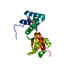

 Mass: 363.221 Da / Num. of mol.: 2 / Source method: obtained synthetically / Formula: C10H14N5O8P / Feature type: SUBJECT OF INVESTIGATION
 HSQC
HSQC Sample preparation
 Processing