- PDB-8pai: Crystal structure of human Histidine Triad Nucleotide-Binding Pro... -
+
Open data
ID or keywords:
Loading...
-
Basic information
Entry
Database: PDB / ID: 8pai
Title
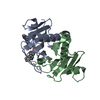
Crystal structure of human Histidine Triad Nucleotide-Binding Protein 1 in complex with 5'-O-[N-(3-Indolepropionic acid)sulfamoyl] N2-methyl-2-aminoethenoadenosine
Components
Histidine triad nucleotide-binding protein 1
Keywords
HYDROLASE / HINT / HIT / histidine triad / phosphoramidase / complex / inhibitor
Function / homology
Function and homology information
purine ribonucleotide catabolic process / Hydrolases; Acting on phosphorus-nitrogen bonds / adenosine 5'-monophosphoramidase activity / deSUMOylase activity / protein desumoylation / Regulation of MITF-M-dependent genes involved in apoptosis / histone deacetylase complex / intrinsic apoptotic signaling pathway by p53 class mediator / Regulation of MITF-M-dependent genes involved in cell cycle and proliferation / Transcriptional and post-translational regulation of MITF-M expression and activity ...purine ribonucleotide catabolic process / Hydrolases; Acting on phosphorus-nitrogen bonds / adenosine 5'-monophosphoramidase activity / deSUMOylase activity / protein desumoylation / Regulation of MITF-M-dependent genes involved in apoptosis / histone deacetylase complex / intrinsic apoptotic signaling pathway by p53 class mediator / Regulation of MITF-M-dependent genes involved in cell cycle and proliferation / Transcriptional and post-translational regulation of MITF-M expression and activity / positive regulation of calcium-mediated signaling / protein kinase C binding / cytoskeleton / Hydrolases; Acting on peptide bonds (peptidases); Cysteine endopeptidases / nucleotide binding / hydrolase activity / regulation of DNA-templated transcription / signal transduction / proteolysis / extracellular exosome / nucleoplasm / nucleus / plasma membrane / cytoplasm / cytosol Similarity search - Function
Histidine triad (HIT) protein / HIT domain / Histidine triad, conserved site / HIT domain signature. / HIT domain profile. / HIT-like domain / HIT-like superfamily Similarity search - Domain/homology
Histidinetriadnucleotide-bindingprotein1 / Adenosine 5'-monophosphoramidase / Protein kinase C inhibitor 1 / Protein kinase C-interacting ...Adenosine 5'-monophosphoramidase / Protein kinase C inhibitor 1 / Protein kinase C-interacting protein 1 / PKCI-1
Mass: 13823.931 Da / Num. of mol.: 2 Source method: isolated from a genetically manipulated source Source: (gene. exp.) Homo sapiens (human) / Gene: HINT1, HINT, PKCI1, PRKCNH1 / Plasmid: pSGA02 / Production host: Escherichia coli BL21(DE3) (bacteria) / References: UniProt: P49773, Hydrolases
Method to determine structure: MOLECULAR REPLACEMENT / Resolution: 1.8→19.041 Å / Cor.coef. Fo:Fc: 0.964 / Cor.coef. Fo:Fc free: 0.946 / SU B: 2.504 / SU ML: 0.078 / Cross valid method: THROUGHOUT / ESU R: 0.12 / ESU R Free: 0.115 Details: Hydrogens have been added in their riding positions
Rfactor
Num. reflection
% reflection
Rfree
0.1906
982
4.651 %
Rwork
0.1494
20130
-
all
0.151
-
-
obs
-
21112
99.688 %
Solvent computation
Ion probe radii: 0.8 Å / Shrinkage radii: 0.8 Å / VDW probe radii: 1.2 Å / Solvent model: MASK BULK SOLVENT
Displacement parameters
Biso mean: 13.439 Å2
Baniso -1
Baniso -2
Baniso -3
1-
1.55 Å2
0 Å2
-0.247 Å2
2-
-
-0.729 Å2
0 Å2
3-
-
-
-0.771 Å2
Refinement step
Cycle: LAST / Resolution: 1.8→19.041 Å
Protein
Nucleic acid
Ligand
Solvent
Total
Num. atoms
1767
0
40
238
2045
Refine LS restraints
Refine-ID
Type
Dev ideal
Dev ideal target
Number
X-RAY DIFFRACTION
r_bond_refined_d
0.008
0.012
2006
X-RAY DIFFRACTION
r_bond_other_d
0.001
0.016
1878
X-RAY DIFFRACTION
r_angle_refined_deg
1.528
1.658
2737
X-RAY DIFFRACTION
r_angle_other_deg
0.531
1.602
4368
X-RAY DIFFRACTION
r_dihedral_angle_1_deg
6.63
5
257
X-RAY DIFFRACTION
r_dihedral_angle_2_deg
5.732
5
12
X-RAY DIFFRACTION
r_dihedral_angle_3_deg
14.207
10
344
X-RAY DIFFRACTION
r_dihedral_angle_6_deg
16.498
10
86
X-RAY DIFFRACTION
r_chiral_restr
0.078
0.2
286
X-RAY DIFFRACTION
r_gen_planes_refined
0.008
0.02
2377
X-RAY DIFFRACTION
r_gen_planes_other
0.002
0.02
432
X-RAY DIFFRACTION
r_nbd_refined
0.224
0.2
419
X-RAY DIFFRACTION
r_symmetry_nbd_other
0.196
0.2
1795
X-RAY DIFFRACTION
r_nbtor_refined
0.173
0.2
976
X-RAY DIFFRACTION
r_symmetry_nbtor_other
0.08
0.2
1067
X-RAY DIFFRACTION
r_xyhbond_nbd_refined
0.174
0.2
155
X-RAY DIFFRACTION
r_symmetry_nbd_refined
0.205
0.2
15
X-RAY DIFFRACTION
r_nbd_other
0.146
0.2
52
X-RAY DIFFRACTION
r_symmetry_xyhbond_nbd_refined
0.201
0.2
15
X-RAY DIFFRACTION
r_mcbond_it
1.259
1.235
989
X-RAY DIFFRACTION
r_mcbond_other
1.256
1.234
988
X-RAY DIFFRACTION
r_mcangle_it
1.973
2.213
1259
X-RAY DIFFRACTION
r_mcangle_other
1.972
2.212
1260
X-RAY DIFFRACTION
r_scbond_it
2.205
1.557
1017
X-RAY DIFFRACTION
r_scbond_other
2.205
1.557
1017
X-RAY DIFFRACTION
r_scangle_it
3.332
2.756
1478
X-RAY DIFFRACTION
r_scangle_other
3.33
2.756
1479
X-RAY DIFFRACTION
r_lrange_it
5.811
15.242
2366
X-RAY DIFFRACTION
r_lrange_other
5.598
14.018
2317
LS refinement shell
Refine-ID: X-RAY DIFFRACTION / Total num. of bins used: 20
Resolution (Å)
Rfactor Rfree
Num. reflection Rfree
Rfactor Rwork
Num. reflection Rwork
Rfactor all
Num. reflection all
Fsc free
Fsc work
% reflection obs (%)
WRfactor Rwork
1.8-1.846
0.223
78
0.192
1443
0.194
1524
0.97
0.975
99.8031
0.17
1.846-1.896
0.259
68
0.175
1431
0.179
1500
0.961
0.977
99.9333
0.149
1.896-1.951
0.204
81
0.16
1388
0.162
1470
0.975
0.983
99.932
0.136
1.951-2.01
0.183
77
0.157
1338
0.159
1415
0.976
0.983
100
0.134
2.01-2.075
0.227
68
0.152
1296
0.156
1365
0.967
0.985
99.9267
0.132
2.075-2.147
0.205
64
0.152
1293
0.155
1358
0.978
0.986
99.9264
0.132
2.147-2.227
0.165
59
0.137
1221
0.138
1280
0.983
0.988
100
0.122
2.227-2.317
0.193
59
0.131
1189
0.134
1250
0.978
0.989
99.84
0.115
2.317-2.418
0.166
59
0.128
1137
0.13
1197
0.981
0.99
99.9165
0.114
2.418-2.534
0.17
48
0.138
1089
0.14
1137
0.981
0.988
100
0.12
2.534-2.669
0.195
53
0.142
1035
0.144
1089
0.98
0.988
99.9082
0.125
2.669-2.827
0.195
45
0.136
991
0.139
1036
0.98
0.989
100
0.121
2.827-3.018
0.251
36
0.137
945
0.141
981
0.963
0.988
100
0.128
3.018-3.253
0.155
35
0.141
873
0.141
908
0.985
0.988
100
0.133
3.253-3.553
0.175
28
0.145
799
0.146
833
0.983
0.987
99.2797
0.138
3.553-3.955
0.151
33
0.146
731
0.146
766
0.986
0.988
99.7389
0.145
3.955-4.534
0.161
26
0.128
657
0.13
690
0.989
0.99
98.9855
0.128
4.534-5.476
0.171
24
0.169
550
0.169
580
0.98
0.986
98.9655
0.166
5.476-7.443
0.236
36
0.219
438
0.221
474
0.967
0.976
100
0.218
7.443-19.041
0.274
5
0.189
286
0.191
302
0.984
0.981
96.3576
0.198
+
About Yorodumi
-
News
-
Feb 9, 2022. New format data for meta-information of EMDB entries
New format data for meta-information of EMDB entries
Version 3 of the EMDB header file is now the official format.
The previous official version 1.9 will be removed from the archive.
In the structure databanks used in Yorodumi, some data are registered as the other names, "COVID-19 virus" and "2019-nCoV". Here are the details of the virus and the list of structure data.
Jan 31, 2019. EMDB accession codes are about to change! (news from PDBe EMDB page)
EMDB accession codes are about to change! (news from PDBe EMDB page)
The allocation of 4 digits for EMDB accession codes will soon come to an end. Whilst these codes will remain in use, new EMDB accession codes will include an additional digit and will expand incrementally as the available range of codes is exhausted. The current 4-digit format prefixed with “EMD-” (i.e. EMD-XXXX) will advance to a 5-digit format (i.e. EMD-XXXXX), and so on. It is currently estimated that the 4-digit codes will be depleted around Spring 2019, at which point the 5-digit format will come into force.
The EM Navigator/Yorodumi systems omit the EMD- prefix.
Related info.:Q: What is EMD? / ID/Accession-code notation in Yorodumi/EM Navigator
Yorodumi is a browser for structure data from EMDB, PDB, SASBDB, etc.
This page is also the successor to EM Navigator detail page, and also detail information page/front-end page for Omokage search.
The word "yorodu" (or yorozu) is an old Japanese word meaning "ten thousand". "mi" (miru) is to see.
Related info.:EMDB / PDB / SASBDB / Comparison of 3 databanks / Yorodumi Search / Aug 31, 2016. New EM Navigator & Yorodumi / Yorodumi Papers / Jmol/JSmol / Function and homology information / Changes in new EM Navigator and Yorodumi
 Movie
Movie Controller

 Yorodumi
 Open data
 Basic information
 Components
 Keywords
 Function and homology information
 Homo sapiens (human)
 X-RAY DIFFRACTION /
  Authors
 United States, 1items
  Citation
 Structure visualization
 Downloads & links
 Other downloads


 PDBj
PDBj


 Assembly
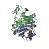

 Mass: 570.578 Da / Num. of mol.: 1 / Source method: obtained synthetically / Formula: C24H26N8O7S / Feature type: SUBJECT OF INVESTIGATION
 Mass: 18.015 Da / Num. of mol.: 238 / Source method: isolated from a natural source / Formula: H2O
 Sample preparation
 Processing