 Movie
Movie Controller


+ Open data

- Basic information
| Entry | Database: PDB / ID: 8p1x | ||||||
|---|---|---|---|---|---|---|---|
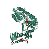
| Title | TarM(Se)_G117R-UDP-glucose | ||||||
 Components Components | TarM(Se) | ||||||
 Keywords Keywords | TRANSFERASE / Staphylococcus epidermidis / glycosyltransferase / GT-B fold / alpha-O-glucose / wall teichoic acid | ||||||
| Function / homology | TRIETHYLENE GLYCOL / PHOSPHATE ION / URIDINE-5'-DIPHOSPHATE-GLUCOSE Function and homology information Function and homology information | ||||||
| Biological species |  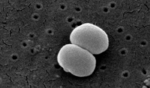 Staphylococcus epidermidis (bacteria) Staphylococcus epidermidis (bacteria) | ||||||
| Method |  X-RAY DIFFRACTION / SYNCHROTRON / MIR / Resolution: 2.03 Å X-RAY DIFFRACTION / SYNCHROTRON / MIR / Resolution: 2.03 Å | ||||||
 Authors Authors | Guo, Y. / Stehle, T. | ||||||
| Funding support |  Germany, 1items Germany, 1items
| ||||||
 Citation Citation | Journal: Sci Adv / Year: 2023 Title: Invasive Staphylococcus epidermidis uses a unique processive wall teichoic acid glycosyltransferase to evade immune recognition. Authors: Guo, Y. / Du, X. / Krusche, J. / Beck, C. / Ali, S. / Walter, A. / Winstel, V. / Mayer, C. / Codee, J.D.C. / Peschel, A. / Stehle, T. | ||||||
| History |
|
- Structure visualization
| Structure viewer | Molecule: MolmilJmol/JSmol |
|---|
- Downloads & links
-Download
| PDBx/mmCIF format | 8p1x.cif.gz | 128.4 KB | Display | PDBx/mmCIF format |
|---|---|---|---|---|
| PDB format | pdb8p1x.ent.gz | Display | PDB format | |
| PDBx/mmJSON format | 8p1x.json.gz | Tree view | PDBx/mmJSON format | |
| Others |  Other downloads Other downloads |
-Validation report
| Arichive directory | https://data.pdbj.org/pub/pdb/validation_reports/p1/8p1xftp://data.pdbj.org/pub/pdb/validation_reports/p1/8p1x | HTTPS FTP |
|---|
-Related structure data


-Links
 PDBj
PDBj
- Assembly
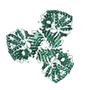
| Deposited unit | 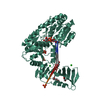
| ||||||||
|---|---|---|---|---|---|---|---|---|---|
| 1 |
| ||||||||
| Unit cell |
|
-Components
-Protein , 1 types, 1 molecules AAA
| #1: Protein | Mass: 59652.496 Da / Num. of mol.: 1 Source method: isolated from a genetically manipulated source Source: (gene. exp.) Staphylococcus epidermidis (bacteria) / Production host: |
|---|
-Non-polymers , 7 types, 300 molecules 


| #2: Chemical |  Mass: 35.453 Da / Num. of mol.: 3 / Source method: obtained synthetically / Formula: Cl Mass: 35.453 Da / Num. of mol.: 3 / Source method: obtained synthetically / Formula: Cl#3: Chemical |  Mass: 94.971 Da / Num. of mol.: 3 / Source method: obtained synthetically / Formula: PO4 Mass: 94.971 Da / Num. of mol.: 3 / Source method: obtained synthetically / Formula: PO4#4: Chemical | ChemComp-UPG / |  Mass: 566.302 Da / Num. of mol.: 1 / Source method: obtained synthetically / Formula: C15H24N2O17P2 / Feature type: SUBJECT OF INVESTIGATION Mass: 566.302 Da / Num. of mol.: 1 / Source method: obtained synthetically / Formula: C15H24N2O17P2 / Feature type: SUBJECT OF INVESTIGATION#5: Chemical | ChemComp-EDO / |  Mass: 62.068 Da / Num. of mol.: 1 / Source method: obtained synthetically / Formula: C2H6O2 Mass: 62.068 Da / Num. of mol.: 1 / Source method: obtained synthetically / Formula: C2H6O2#6: Chemical | ChemComp-PG4 / |  Mass: 194.226 Da / Num. of mol.: 1 / Source method: obtained synthetically / Formula: C8H18O5 / Comment: precipitant*YM Mass: 194.226 Da / Num. of mol.: 1 / Source method: obtained synthetically / Formula: C8H18O5 / Comment: precipitant*YM#7: Chemical | ChemComp-PGE / |  Mass: 150.173 Da / Num. of mol.: 1 / Source method: obtained synthetically / Formula: C6H14O4 Mass: 150.173 Da / Num. of mol.: 1 / Source method: obtained synthetically / Formula: C6H14O4#8: Water | ChemComp-HOH / | Mass: 18.015 Da / Num. of mol.: 290 / Source method: isolated from a natural source / Formula: H2O |
|---|
-Details
| Has ligand of interest | Y |
|---|
-Experimental details
-Experiment
| Experiment | Method: X-RAY DIFFRACTION / Number of used crystals: 1 |
|---|
- Sample preparation
| Crystal | Density Matthews: 2.14 Å3/Da / Density % sol: 42.48 % |
|---|---|
| Crystal grow | Temperature: 293 K / Method: vapor diffusion, hanging drop / pH: 6.9 Details: 10% PEG 20 000, 25% PEG MME 550, 0.1 M MES/imidazole, pH 6.9, 0.03 M NaNO3, 0.03 M Na2HPO4, 0.03 M (NH4)2SO4. PH range: 6.2-8.1 |
-Data collection
| Diffraction | Mean temperature: 100 K / Serial crystal experiment: N |
|---|---|
| Diffraction source | Source: SYNCHROTRON / Site: SLS  / Beamline: X06DA / Wavelength: 1 Å / Beamline: X06DA / Wavelength: 1 Å |
| Detector | Type: DECTRIS PILATUS 2M / Detector: PIXEL / Date: Feb 7, 2020 |
| Radiation | Protocol: SINGLE WAVELENGTH / Monochromatic (M) / Laue (L): M / Scattering type: x-ray |
| Radiation wavelength | Wavelength: 1 Å / Relative weight: 1 |
| Reflection | Resolution: 2.03→49.03 Å / Num. obs: 33765 / % possible obs: 100 % / Redundancy: 13.2 % / CC1/2: 0.999 / Net I/σ(I): 15.64 |
| Reflection shell | Resolution: 2.03→2.08 Å / Num. unique obs: 2443 / CC1/2: 0.52 / % possible all: 100 |
- Processing
| Software |
| ||||||||||||||||||||||||||||||||||||||||||||||||||||||||||||||||||||||||||||||||||||||||||||||||||||||||||||||||||||||||||||||||||||||||||||||||||||||
|---|---|---|---|---|---|---|---|---|---|---|---|---|---|---|---|---|---|---|---|---|---|---|---|---|---|---|---|---|---|---|---|---|---|---|---|---|---|---|---|---|---|---|---|---|---|---|---|---|---|---|---|---|---|---|---|---|---|---|---|---|---|---|---|---|---|---|---|---|---|---|---|---|---|---|---|---|---|---|---|---|---|---|---|---|---|---|---|---|---|---|---|---|---|---|---|---|---|---|---|---|---|---|---|---|---|---|---|---|---|---|---|---|---|---|---|---|---|---|---|---|---|---|---|---|---|---|---|---|---|---|---|---|---|---|---|---|---|---|---|---|---|---|---|---|---|---|---|---|---|---|---|
| Refinement | Method to determine structure: MIR / Resolution: 2.03→49.025 Å / Cor.coef. Fo:Fc: 0.953 / Cor.coef. Fo:Fc free: 0.937 / SU B: 3.068 / SU ML: 0.089 / Cross valid method: FREE R-VALUE / ESU R: 0.046 / ESU R Free: 0.038 Details: Hydrogens have been added in their riding positions
| ||||||||||||||||||||||||||||||||||||||||||||||||||||||||||||||||||||||||||||||||||||||||||||||||||||||||||||||||||||||||||||||||||||||||||||||||||||||
| Solvent computation | Ion probe radii: 0.8 Å / Shrinkage radii: 0.8 Å / VDW probe radii: 1.2 Å / Solvent model: MASK BULK SOLVENT | ||||||||||||||||||||||||||||||||||||||||||||||||||||||||||||||||||||||||||||||||||||||||||||||||||||||||||||||||||||||||||||||||||||||||||||||||||||||
| Displacement parameters | Biso mean: 34.271 Å2
| ||||||||||||||||||||||||||||||||||||||||||||||||||||||||||||||||||||||||||||||||||||||||||||||||||||||||||||||||||||||||||||||||||||||||||||||||||||||
| Refinement step | Cycle: LAST / Resolution: 2.03→49.025 Å
| ||||||||||||||||||||||||||||||||||||||||||||||||||||||||||||||||||||||||||||||||||||||||||||||||||||||||||||||||||||||||||||||||||||||||||||||||||||||
| Refine LS restraints |
| ||||||||||||||||||||||||||||||||||||||||||||||||||||||||||||||||||||||||||||||||||||||||||||||||||||||||||||||||||||||||||||||||||||||||||||||||||||||
| LS refinement shell |
|
