[English] 日本語
Yorodumi
- PDB-8oyk: Coiled-Coil Domain of Human STIL, L736E Mutant -

+
Open data


ID or keywords:

Loading...

-
Basic information

Entry
Database: PDB / ID: 8oyk
TitleCoiled-Coil Domain of Human STIL, L736E Mutant
ComponentsIsoform 2 of SCL-interrupting locus protein
KeywordsSTRUCTURAL PROTEIN / STIL / coiled coil / dimer / antiparallel
Function / homology
Function and homology information


floor plate development / regulation of centriole replication / procentriole replication complex / positive regulation of centriole replication / embryonic axis specification / positive regulation of spindle assembly / notochord development / determination of left/right symmetry / protein localization to centrosome / neural tube development ...floor plate development / regulation of centriole replication / procentriole replication complex / positive regulation of centriole replication / embryonic axis specification / positive regulation of spindle assembly / notochord development / determination of left/right symmetry / protein localization to centrosome / neural tube development / smoothened signaling pathway / heart looping / centrosome duplication / positive regulation of G1/S transition of mitotic cell cycle / regulation of mitotic spindle organization / forebrain development / centriole / mitotic spindle organization / neural tube closure / multicellular organism growth / cell cortex / in utero embryonic development / centrosome / negative regulation of apoptotic process / nucleoplasm / identical protein binding / cytoplasm / cytosol
Similarity search - Function
SCL-interrupting locus protein / SCL-interrupting locus protein N-terminus
Similarity search - Domain/homology
SCL-interrupting locus protein
Similarity search - Component
Biological speciesHomo sapiens (human)
MethodX-RAY DIFFRACTION / SYNCHROTRON / AB INITIO PHASING / Resolution: 1.9 Å
AuthorsMartin, F.J.O. / Shamir, M. / Woolfson, D.N. / Friedler, A.
Funding supportEuropean Union, United Kingdom, Israel, 4items
OrganizationGrant numberCountry
European Research Council (ERC)340764European Union
Engineering and Physical Sciences Research CouncilEP/G036764 United Kingdom
Biotechnology and Biological Sciences Research Council (BBSRC)BB R00661X 1 United Kingdom
Israel Science Foundation939/14 Israel
CitationJournal: Int J Mol Sci / Year: 2023
Title: Molecular Mechanism of STIL Coiled-Coil Domain Oligomerization.
Authors: Shamir, M. / Martin, F.J.O. / Woolfson, D.N. / Friedler, A.
History
DepositionMay 5, 2023Deposition site: PDBE / Processing site: PDBE
SupersessionOct 18, 2023ID: 7QXG
Revision 1.0Oct 18, 2023Provider: repository / Type: Initial release
Revision 1.1Oct 25, 2023Group: Database references / Category: citation
Item: _citation.journal_volume / _citation.pdbx_database_id_PubMed / _citation.title

-
Structure visualization

Structure viewerMolecule:
MolmilJmol/JSmol

Downloads & links

-
Assembly

Deposited unit
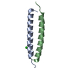
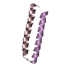
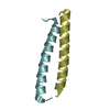
A: Isoform 2 of SCL-interrupting locus protein
B: Isoform 2 of SCL-interrupting locus protein
C: Isoform 2 of SCL-interrupting locus protein
D: Isoform 2 of SCL-interrupting locus protein
E: Isoform 2 of SCL-interrupting locus protein
F: Isoform 2 of SCL-interrupting locus protein
hetero molecules


Theoretical massNumber of molelcules
Total (without water)23,0417
Polymers23,0066
Non-polymers351
Water1,892105
1
A: Isoform 2 of SCL-interrupting locus protein
B: Isoform 2 of SCL-interrupting locus protein
hetero molecules


Theoretical massNumber of molelcules
Total (without water)7,7043
Polymers7,6692
Non-polymers351
Water362
TypeNameSymmetry operationNumber
identity operation1_555x,y,z1
Buried area1480 Å2
ΔGint-18 kcal/mol
Surface area5480 Å2
2
C: Isoform 2 of SCL-interrupting locus protein
E: Isoform 2 of SCL-interrupting locus protein


Theoretical massNumber of molelcules
Total (without water)7,6692
Polymers7,6692
Non-polymers00
Water362
TypeNameSymmetry operationNumber
identity operation1_555x,y,z1
Buried area1290 Å2
ΔGint-13 kcal/mol
Surface area4790 Å2
3
D: Isoform 2 of SCL-interrupting locus protein
F: Isoform 2 of SCL-interrupting locus protein


Theoretical massNumber of molelcules
Total (without water)7,6692
Polymers7,6692
Non-polymers00
Water362
TypeNameSymmetry operationNumber
identity operation1_555x,y,z1
Buried area1420 Å2
ΔGint-15 kcal/mol
Surface area5020 Å2
Unit cell
Length a, b, c (Å)43.539, 31.269, 64.473
Angle α, β, γ (deg.)90.00, 98.08, 90.00
Int Tables number4
Space group name H-MP1211

-
Components

#1: Protein/peptide
Isoform 2 of SCL-interrupting locus protein / TAL-1-interrupting locus protein


Mass: 3834.298 Da / Num. of mol.: 6 / Source method: obtained synthetically / Source: (synth.) Homo sapiens (human) / References: UniProt: Q15468
#2: Chemical ChemComp-CL / CHLORIDE ION


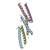
Mass: 35.453 Da / Num. of mol.: 1 / Source method: obtained synthetically / Formula: Cl
#3: Water ChemComp-HOH / water


Mass: 18.015 Da / Num. of mol.: 105 / Source method: isolated from a natural source / Formula: H2O
Has ligand of interestN

-
Experimental details

-
Experiment

ExperimentMethod: X-RAY DIFFRACTION / Number of used crystals: 1

-
Sample preparation

CrystalDensity Matthews: 1.89 Å3/Da / Density % sol: 34.88 %
Crystal growTemperature: 277 K / Method: vapor diffusion / pH: 8
Details: 0.9 mM peptide, 50 mM Tris, and 10 % v/v MPD, at pH 8.0.

-
Data collection

DiffractionMean temperature: 100 K / Serial crystal experiment: N
Diffraction sourceSource: SYNCHROTRON / Site: Diamond / Beamline: I24 / Wavelength: 0.97 Å
DetectorType: DECTRIS PILATUS3 6M / Detector: PIXEL / Date: Jun 29, 2019
RadiationProtocol: SINGLE WAVELENGTH / Monochromatic (M) / Laue (L): M / Scattering type: x-ray
Radiation wavelengthWavelength: 0.97 Å / Relative weight: 1
ReflectionResolution: 1.9→63.83 Å / Num. obs: 13477 / % possible obs: 97.7 % / Redundancy: 1.9 % / Rmerge(I) obs: 0.038 / Rpim(I) all: 0.038 / Rrim(I) all: 0.054 / Net I/σ(I): 15.4
Reflection shellResolution: 1.9→1.94 Å / Redundancy: 1.9 % / Rmerge(I) obs: 0.119 / Mean I/σ(I) obs: 6 / Num. unique obs: 846 / CC1/2: 0.957 / Rpim(I) all: 0.119 / Rrim(I) all: 0.168 / % possible all: 96.6

-
Processing

Software
NameVersionClassification
REFMAC5.8.0411refinement
autoPROC2.2.19data reduction
Aimless0.7.7data scaling
Arcimboldo1.0.21phasing
RefinementMethod to determine structure: AB INITIO PHASING / Resolution: 1.9→63.83 Å / Cor.coef. Fo:Fc: 0.947 / Cor.coef. Fo:Fc free: 0.922 / SU B: 7.311 / SU ML: 0.108 / Cross valid method: THROUGHOUT / ESU R: 0.187 / ESU R Free: 0.16 / Stereochemistry target values: MAXIMUM LIKELIHOOD / Details: HYDROGENS HAVE BEEN ADDED IN THE RIDING POSITIONS
RfactorNum. reflection% reflectionSelection details
Rfree0.22492 680 5 %RANDOM
Rwork0.18048 ---
obs0.18274 12795 97.11 %-
Solvent computationIon probe radii: 0.8 Å / Shrinkage radii: 0.8 Å / VDW probe radii: 1.1 Å / Solvent model: MASK
Displacement parametersBiso mean: 20.465 Å2
Baniso -1Baniso -2Baniso -3
1-0.34 Å20 Å2-0.29 Å2
2--0.23 Å20 Å2
3----0.47 Å2
Refinement stepCycle: 1 / Resolution: 1.9→63.83 Å
ProteinNucleic acidLigandSolventTotal
Num. atoms1549 0 1 105 1655
Refine LS restraints
Refine-IDTypeDev idealDev ideal targetNumber
X-RAY DIFFRACTIONr_bond_refined_d0.0080.0121609
X-RAY DIFFRACTIONr_bond_other_d0.0010.0161597
X-RAY DIFFRACTIONr_angle_refined_deg1.3541.6542173
X-RAY DIFFRACTIONr_angle_other_deg0.4631.5673633
X-RAY DIFFRACTIONr_dihedral_angle_1_deg4.6035190
X-RAY DIFFRACTIONr_dihedral_angle_2_deg5.166526
X-RAY DIFFRACTIONr_dihedral_angle_3_deg14.17410315
X-RAY DIFFRACTIONr_dihedral_angle_4_deg
X-RAY DIFFRACTIONr_chiral_restr0.0690.2241
X-RAY DIFFRACTIONr_gen_planes_refined0.0070.022024
X-RAY DIFFRACTIONr_gen_planes_other0.0010.02432
X-RAY DIFFRACTIONr_nbd_refined
X-RAY DIFFRACTIONr_nbd_other
X-RAY DIFFRACTIONr_nbtor_refined
X-RAY DIFFRACTIONr_nbtor_other
X-RAY DIFFRACTIONr_xyhbond_nbd_refined
X-RAY DIFFRACTIONr_xyhbond_nbd_other
X-RAY DIFFRACTIONr_metal_ion_refined
X-RAY DIFFRACTIONr_metal_ion_other
X-RAY DIFFRACTIONr_symmetry_vdw_refined
X-RAY DIFFRACTIONr_symmetry_vdw_other
X-RAY DIFFRACTIONr_symmetry_hbond_refined
X-RAY DIFFRACTIONr_symmetry_hbond_other
X-RAY DIFFRACTIONr_symmetry_metal_ion_refined
X-RAY DIFFRACTIONr_symmetry_metal_ion_other
X-RAY DIFFRACTIONr_mcbond_it2.7720.955743
X-RAY DIFFRACTIONr_mcbond_other2.6720.955743
X-RAY DIFFRACTIONr_mcangle_it3.2091.679923
X-RAY DIFFRACTIONr_mcangle_other3.2351.683924
X-RAY DIFFRACTIONr_scbond_it6.231.538866
X-RAY DIFFRACTIONr_scbond_other6.2261.539867
X-RAY DIFFRACTIONr_scangle_it
X-RAY DIFFRACTIONr_scangle_other7.9312.5521245
X-RAY DIFFRACTIONr_long_range_B_refined8.62215.121890
X-RAY DIFFRACTIONr_long_range_B_other8.58214.671870
X-RAY DIFFRACTIONr_rigid_bond_restr
X-RAY DIFFRACTIONr_sphericity_free
X-RAY DIFFRACTIONr_sphericity_bonded
LS refinement shellResolution: 1.9→1.949 Å / Total num. of bins used: 20
RfactorNum. reflection% reflection
Rfree0.254 58 -
Rwork0.203 912 -
obs--96.23 %
Refinement TLS params.

Method: refined / Refine-ID: X-RAY DIFFRACTION

IDL112)L122)L132)L222)L232)L332)S11 (Å °)S12 (Å °)S13 (Å °)S21 (Å °)S22 (Å °)S23 (Å °)S31 (Å °)S32 (Å °)S33 (Å °)T112)T122)T132)T222)T232)T332)Origin x (Å)Origin y (Å)Origin z (Å)
11.34630.25412.07160.9271.36577.396-0.00320.0339-0.034-0.0210.0135-0.018-0.03930.0927-0.01030.02-0.00910.00850.01560.01740.049814.56145.779215.9899
27.47613.14239.20571.87154.780513.2305-0.1567-0.10090.29-0.02210.01070.0949-0.1452-0.06890.1460.00830.00850.00250.00940.00760.083712.754713.702516.513
31.6365-0.13352.96180.664-0.83069.31420.01960.0176-0.06380.00220.03120.0310.06840.0016-0.05080.0320.00740.01680.0099-0.01230.056731.21143.503435.6212
411.6851-0.829511.34253.2681-2.180812.77190.03260.0010.07510.06-0.09740.0143-0.19-0.05440.06480.04020.00930.00950.0147-0.00730.0127-2.49240.05176.6229
54.5619-2.94166.32413.0377-4.473310.505-0.05010.07860.0561-0.05090.0126-0.12430.04460.030.03740.0169-0.01350.01310.0113-0.0110.031734.971510.765528.1585
610.03813.159311.73942.47213.079316.47590.1697-0.42950.02570.1019-0.09030.17560.0891-0.3122-0.07930.05920.02610.03780.1086-0.02580.097-11.8396-0.97628.6591
Refinement TLS group
IDRefine-IDRefine TLS-IDAuth asym-IDAuth seq-ID
1X-RAY DIFFRACTION1A1 - 32
2X-RAY DIFFRACTION2B1 - 32
3X-RAY DIFFRACTION3C1 - 31
4X-RAY DIFFRACTION4D1 - 29
5X-RAY DIFFRACTION5E1 - 30
6X-RAY DIFFRACTION6F1 - 32

+
About Yorodumi

-
News

-
Feb 9, 2022. New format data for meta-information of EMDB entries

New format data for meta-information of EMDB entries

  • Version 3 of the EMDB header file is now the official format.
  • The previous official version 1.9 will be removed from the archive.

Related info.:EMDB header

External links:wwPDB to switch to version 3 of the EMDB data model

-
Aug 12, 2020. Covid-19 info

Covid-19 info

URL: https://pdbjlvh1.pdbj.org/emnavi/covid19.php

New page: Covid-19 featured information page in EM Navigator.

Related info.:Covid-19 info / Mar 5, 2020. Novel coronavirus structure data

+
Mar 5, 2020. Novel coronavirus structure data

Novel coronavirus structure data

Related info.:Yorodumi Speices / Aug 12, 2020. Covid-19 info

External links:COVID-19 featured content - PDBj / Molecule of the Month (242):Coronavirus Proteases

+
Jan 31, 2019. EMDB accession codes are about to change! (news from PDBe EMDB page)

EMDB accession codes are about to change! (news from PDBe EMDB page)

  • The allocation of 4 digits for EMDB accession codes will soon come to an end. Whilst these codes will remain in use, new EMDB accession codes will include an additional digit and will expand incrementally as the available range of codes is exhausted. The current 4-digit format prefixed with “EMD-” (i.e. EMD-XXXX) will advance to a 5-digit format (i.e. EMD-XXXXX), and so on. It is currently estimated that the 4-digit codes will be depleted around Spring 2019, at which point the 5-digit format will come into force.
  • The EM Navigator/Yorodumi systems omit the EMD- prefix.

Related info.:Q: What is EMD? / ID/Accession-code notation in Yorodumi/EM Navigator

External links:EMDB Accession Codes are Changing Soon! / Contact to PDBj

+
Jul 12, 2017. Major update of PDB

Major update of PDB

  • wwPDB released updated PDB data conforming to the new PDBx/mmCIF dictionary.
  • This is a major update changing the version number from 4 to 5, and with Remediation, in which all the entries are updated.
  • In this update, many items about electron microscopy experimental information are reorganized (e.g. em_software).
  • Now, EM Navigator and Yorodumi are based on the updated data.

External links:wwPDB Remediation / Enriched Model Files Conforming to OneDep Data Standards Now Available in the PDB FTP Archive

-
Yorodumi

Thousand views of thousand structures

  • Yorodumi is a browser for structure data from EMDB, PDB, SASBDB, etc.
  • This page is also the successor to EM Navigator detail page, and also detail information page/front-end page for Omokage search.
  • The word "yorodu" (or yorozu) is an old Japanese word meaning "ten thousand". "mi" (miru) is to see.

Related info.:EMDB / PDB / SASBDB / Comparison of 3 databanks / Yorodumi Search / Aug 31, 2016. New EM Navigator & Yorodumi / Yorodumi Papers / Jmol/JSmol / Function and homology information / Changes in new EM Navigator and Yorodumi

Read more