 Movie
Movie Controller

[English] 日本語
 Yorodumi
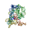
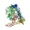
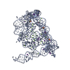
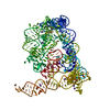
- PDB-8oly: Structure of Oceanobacillus iheyensis group II intron post first ... -
+ Open data

- Basic information
| Entry | Database: PDB / ID: 8oly | |||||||||||||||||||||||||||
|---|---|---|---|---|---|---|---|---|---|---|---|---|---|---|---|---|---|---|---|---|---|---|---|---|---|---|---|---|
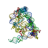
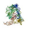
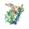
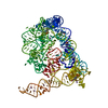
| Title | Structure of Oceanobacillus iheyensis group II intron post first step of splicing in the presence of K+, Mg2+ and intronistat B | |||||||||||||||||||||||||||
 Components Components |
| |||||||||||||||||||||||||||
 Keywords Keywords | RNA / RIBOZYME / METALLOENZYME / SELF-SPLICING / RETROTRANSPOSITION | |||||||||||||||||||||||||||
| Function / homology | : / SPERMINE / RNA / RNA (> 10) / RNA (> 100) Function and homology information Function and homology information | |||||||||||||||||||||||||||
| Biological species |  Oceanobacillus iheyensis (bacteria) Oceanobacillus iheyensis (bacteria) | |||||||||||||||||||||||||||
| Method |  X-RAY DIFFRACTION / SYNCHROTRON / MOLECULAR REPLACEMENT / Resolution: 3.11 Å X-RAY DIFFRACTION / SYNCHROTRON / MOLECULAR REPLACEMENT / Resolution: 3.11 Å | |||||||||||||||||||||||||||
 Authors Authors | Silvestri, I. / Marcia, M. | |||||||||||||||||||||||||||
| Funding support |  France, France,  Italy, 8items Italy, 8items
| |||||||||||||||||||||||||||
 Citation Citation | Journal: Nat Commun / Year: 2024 Title: Targeting the conserved active site of splicing machines with specific and selective small molecule modulators. Authors: Silvestri, I. / Manigrasso, J. / Andreani, A. / Brindani, N. / Mas, C. / Reiser, J.B. / Vidossich, P. / Martino, G. / McCarthy, A.A. / De Vivo, M. / Marcia, M. | |||||||||||||||||||||||||||
| History |
|
- Structure visualization
| Structure viewer | Molecule: MolmilJmol/JSmol |
|---|
- Downloads & links
-Download
| PDBx/mmCIF format | 8oly.cif.gz | 239.6 KB | Display | PDBx/mmCIF format |
|---|---|---|---|---|
| PDB format | pdb8oly.ent.gz | 178.7 KB | Display | PDB format |
| PDBx/mmJSON format | 8oly.json.gz | Tree view | PDBx/mmJSON format | |
| Others |  Other downloads Other downloads |
-Validation report
| Arichive directory | https://data.pdbj.org/pub/pdb/validation_reports/ol/8olyftp://data.pdbj.org/pub/pdb/validation_reports/ol/8oly | HTTPS FTP |
|---|
-Related structure data
| Related structure data | 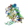 8olsC 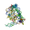 8olvC 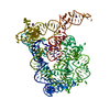 8olwC 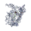 8olzC 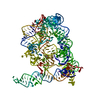 8om0C 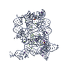 8ruhC 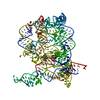 8ruiC  8rujC 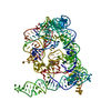 8rukC  8rulC 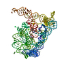 8rumC 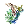 8runC C: citing same article ( |
|---|---|
| Similar structure data |


-Links
 PDBj
PDBj


- Assembly
| Deposited unit | 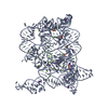
| ||||||||
|---|---|---|---|---|---|---|---|---|---|
| 1 |
| ||||||||
| Unit cell |
|
-Components
-RNA chain , 2 types, 2 molecules AB
| #1: RNA chain | Mass: 126956.180 Da / Num. of mol.: 1 / Source method: obtained synthetically / Source: (synth.) Oceanobacillus iheyensis (bacteria) |
|---|---|
| #2: RNA chain | Mass: 1547.952 Da / Num. of mol.: 1 / Source method: obtained synthetically / Source: (synth.) Oceanobacillus iheyensis (bacteria) |
-Non-polymers , 5 types, 105 molecules 


| #3: Chemical | ChemComp-MG /  Mass: 24.305 Da / Num. of mol.: 30 / Source method: obtained synthetically / Formula: Mg Mass: 24.305 Da / Num. of mol.: 30 / Source method: obtained synthetically / Formula: Mg#4: Chemical | ChemComp-K /  Mass: 39.098 Da / Num. of mol.: 19 / Source method: obtained synthetically / Formula: K Mass: 39.098 Da / Num. of mol.: 19 / Source method: obtained synthetically / Formula: K#5: Chemical | ChemComp-EPE /  Mass: 238.305 Da / Num. of mol.: 5 / Source method: obtained synthetically / Formula: C8H18N2O4S / Comment: pH buffer*YM Mass: 238.305 Da / Num. of mol.: 5 / Source method: obtained synthetically / Formula: C8H18N2O4S / Comment: pH buffer*YM#6: Chemical |  Mass: 202.340 Da / Num. of mol.: 2 / Source method: obtained synthetically / Formula: C10H26N4 Mass: 202.340 Da / Num. of mol.: 2 / Source method: obtained synthetically / Formula: C10H26N4#7: Water | ChemComp-HOH / | Mass: 18.015 Da / Num. of mol.: 49 / Source method: isolated from a natural source / Formula: H2O |
|---|
-Details
| Has ligand of interest | N |
|---|
-Experimental details
-Experiment
| Experiment | Method: X-RAY DIFFRACTION / Number of used crystals: 1 |
|---|
- Sample preparation
| Crystal | Density Matthews: 3.63 Å3/Da / Density % sol: 66.15 % |
|---|---|
| Crystal grow | Temperature: 303.15 K / Method: vapor diffusion, hanging drop Details: 100 mM magnesium acetate, 100 mM potassium chloride, 10 mM lithium chloride, 50 mM HEPES sodium, pH 7.0, 3.5% PEG8000 |
-Data collection
| Diffraction | Mean temperature: 100 K / Serial crystal experiment: N |
|---|---|
| Diffraction source | Source: SYNCHROTRON / Site: ESRF / Beamline: MASSIF-1 / Wavelength: 0.96 Å |
| Detector | Type: DECTRIS PILATUS 2M / Detector: PIXEL / Date: Nov 15, 2021 |
| Radiation | Protocol: SINGLE WAVELENGTH / Monochromatic (M) / Laue (L): M / Scattering type: x-ray |
| Radiation wavelength | Wavelength: 0.96 Å / Relative weight: 1 |
| Reflection | Resolution: 3.1→48.02 Å / Num. obs: 34101 / % possible obs: 98.69 % / Redundancy: 4.2 % / CC1/2: 1 / Net I/σ(I): 10.57 |
| Reflection shell | Resolution: 3.1→3.2 Å / Num. unique obs: 3245 / CC1/2: 0.22 |
- Processing
| Software |
| |||||||||||||||||||||||||||||||||||||||||||||||||||||||||||||||||||||||||||||||||||||||||||||||||||||||||||||||||||||||||||||||||||||||||||||||||||||||||||||||||||||||||||||||||||||||||
|---|---|---|---|---|---|---|---|---|---|---|---|---|---|---|---|---|---|---|---|---|---|---|---|---|---|---|---|---|---|---|---|---|---|---|---|---|---|---|---|---|---|---|---|---|---|---|---|---|---|---|---|---|---|---|---|---|---|---|---|---|---|---|---|---|---|---|---|---|---|---|---|---|---|---|---|---|---|---|---|---|---|---|---|---|---|---|---|---|---|---|---|---|---|---|---|---|---|---|---|---|---|---|---|---|---|---|---|---|---|---|---|---|---|---|---|---|---|---|---|---|---|---|---|---|---|---|---|---|---|---|---|---|---|---|---|---|---|---|---|---|---|---|---|---|---|---|---|---|---|---|---|---|---|---|---|---|---|---|---|---|---|---|---|---|---|---|---|---|---|---|---|---|---|---|---|---|---|---|---|---|---|---|---|---|---|---|
| Refinement | Method to determine structure: MOLECULAR REPLACEMENT / Resolution: 3.11→48.02 Å / Cor.coef. Fo:Fc: 0.961 / Cor.coef. Fo:Fc free: 0.956 / SU B: 23.462 / SU ML: 0.375 / Cross valid method: THROUGHOUT / ESU R Free: 0.388 / Stereochemistry target values: MAXIMUM LIKELIHOOD / Details: HYDROGENS HAVE BEEN ADDED IN THE RIDING POSITIONS
| |||||||||||||||||||||||||||||||||||||||||||||||||||||||||||||||||||||||||||||||||||||||||||||||||||||||||||||||||||||||||||||||||||||||||||||||||||||||||||||||||||||||||||||||||||||||||
| Solvent computation | Ion probe radii: 0.8 Å / Shrinkage radii: 0.8 Å / VDW probe radii: 1.2 Å / Solvent model: MASK | |||||||||||||||||||||||||||||||||||||||||||||||||||||||||||||||||||||||||||||||||||||||||||||||||||||||||||||||||||||||||||||||||||||||||||||||||||||||||||||||||||||||||||||||||||||||||
| Displacement parameters |
| |||||||||||||||||||||||||||||||||||||||||||||||||||||||||||||||||||||||||||||||||||||||||||||||||||||||||||||||||||||||||||||||||||||||||||||||||||||||||||||||||||||||||||||||||||||||||
| Refinement step | Cycle: 1 / Resolution: 3.11→48.02 Å
| |||||||||||||||||||||||||||||||||||||||||||||||||||||||||||||||||||||||||||||||||||||||||||||||||||||||||||||||||||||||||||||||||||||||||||||||||||||||||||||||||||||||||||||||||||||||||
| Refine LS restraints |
| |||||||||||||||||||||||||||||||||||||||||||||||||||||||||||||||||||||||||||||||||||||||||||||||||||||||||||||||||||||||||||||||||||||||||||||||||||||||||||||||||||||||||||||||||||||||||
| LS refinement shell | Resolution: 3.111→3.192 Å / Total num. of bins used: 20
|
