 Movie
Movie Controller


+ Open data

- Basic information
| Entry | Database: PDB / ID: 8ok8 | ||||||
|---|---|---|---|---|---|---|---|
| Title | Variant Surface Glycoprotein VSG615 | ||||||
 Components Components | Variant surface glycoprotein 615 | ||||||
 Keywords Keywords | MEMBRANE PROTEIN / Variant surface glycoprotein / Trypanosomiasis / Glycosylation | ||||||
| Function / homology | Trypanosome variant surface glycoprotein, B-type, N-terminal domain / Trypanosomal VSG domain / Trypanosome variant surface glycoprotein, C-terminal / Trypanosome variant surface glycoprotein C-terminal domain / Variant surface glycoprotein C-terminal domain superfamily / plasma membrane / alpha-D-glucopyranose / Variant surface glycoprotein 615 Function and homology information Function and homology information | ||||||
| Biological species |  | ||||||
| Method |  X-RAY DIFFRACTION / SYNCHROTRON / MOLECULAR REPLACEMENT / Resolution: 3.22 Å X-RAY DIFFRACTION / SYNCHROTRON / MOLECULAR REPLACEMENT / Resolution: 3.22 Å | ||||||
 Authors Authors | Zeelen, J.P. / Stebbins, C.E. / Chandra, M. | ||||||
| Funding support | 1items
| ||||||
 Citation Citation | Journal: Plos Negl Trop Dis / Year: 2023 Title: A structural classification of the variant surface glycoproteins of the African trypanosomey. Authors: Dakovic, S. / Zeelen, J.P. / Gkeka, A. / Chandra, M. / van Straaten, M. / Foti, K. / Zhong, J. / Vlachou, E.P. / Aresta-Branco, F. / Verdi, J.P. / Papavasiliou, F.N. / Stebbins, C.E. | ||||||
| History |
|
- Structure visualization
| Structure viewer | Molecule: MolmilJmol/JSmol |
|---|
- Downloads & links
-Download
| PDBx/mmCIF format | 8ok8.cif.gz | 318.6 KB | Display | PDBx/mmCIF format |
|---|---|---|---|---|
| PDB format | pdb8ok8.ent.gz | 259.1 KB | Display | PDB format |
| PDBx/mmJSON format | 8ok8.json.gz | Tree view | PDBx/mmJSON format | |
| Others |  Other downloads Other downloads |
-Validation report
| Arichive directory | https://data.pdbj.org/pub/pdb/validation_reports/ok/8ok8ftp://data.pdbj.org/pub/pdb/validation_reports/ok/8ok8 | HTTPS FTP |
|---|
-Related structure data
| Related structure data | 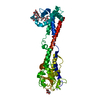 8ok4C  8ok5C 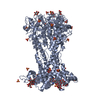 8ok6C  8ok7C 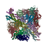 8onhC  8q0eC  8q0pC C: citing same article ( |
|---|---|
| Similar structure data |
-Links
 PDBj
PDBj

- Assembly
| Deposited unit | 
| ||||||||||
|---|---|---|---|---|---|---|---|---|---|---|---|
| 1 | 
| ||||||||||
| 2 | 
| ||||||||||
| Unit cell |
|
-Components
| #1: Protein | Mass: 39941.555 Da / Num. of mol.: 6 Source method: isolated from a genetically manipulated source Source: (gene. exp.) #2: Polysaccharide | Source method: isolated from a genetically manipulated source #3: Sugar | ChemComp-NAG /   Type: D-saccharide, beta linking / Mass: 221.208 Da / Num. of mol.: 4 / Source method: obtained synthetically / Formula: C8H15NO6 / Feature type: SUBJECT OF INVESTIGATION Type: D-saccharide, beta linking / Mass: 221.208 Da / Num. of mol.: 4 / Source method: obtained synthetically / Formula: C8H15NO6 / Feature type: SUBJECT OF INVESTIGATION#4: Sugar | ChemComp-GLC /   Type: D-saccharide, alpha linking / Mass: 180.156 Da / Num. of mol.: 10 / Source method: obtained synthetically / Formula: C6H12O6 / Feature type: SUBJECT OF INVESTIGATION Type: D-saccharide, alpha linking / Mass: 180.156 Da / Num. of mol.: 10 / Source method: obtained synthetically / Formula: C6H12O6 / Feature type: SUBJECT OF INVESTIGATION#5: Water | ChemComp-HOH / |  Mass: 18.015 Da / Num. of mol.: 12 / Source method: isolated from a natural source / Formula: H2O Mass: 18.015 Da / Num. of mol.: 12 / Source method: isolated from a natural source / Formula: H2OHas ligand of interest | Y | Has protein modification | Y | |
|---|
-Experimental details
-Experiment
| Experiment | Method: X-RAY DIFFRACTION / Number of used crystals: 1 |
|---|
- Sample preparation
| Crystal | Density Matthews: 2.3 Å3/Da / Density % sol: 46.42 % |
|---|---|
| Crystal grow | Temperature: 296 K / Method: vapor diffusion, hanging drop / pH: 6 / Details: 23 % PEG 4000, 100 mM NaCacodylate, 10 mM ZnCl2 |
-Data collection
| Diffraction | Mean temperature: 100 K / Serial crystal experiment: N |
|---|---|
| Diffraction source | Source: SYNCHROTRON / Site: SLS  / Beamline: X06DA / Wavelength: 1 Å / Beamline: X06DA / Wavelength: 1 Å |
| Detector | Type: DECTRIS PILATUS 2M-F / Detector: PIXEL / Date: Oct 27, 2019 |
| Radiation | Protocol: SINGLE WAVELENGTH / Monochromatic (M) / Laue (L): M / Scattering type: x-ray |
| Radiation wavelength | Wavelength: 1 Å / Relative weight: 1 |
| Reflection | Resolution: 3.22→77.68 Å / Num. obs: 30929 / % possible obs: 92.38 % / Redundancy: 6 % / Biso Wilson estimate: 85.19 Å2 / CC1/2: 0.978 / Net I/σ(I): 3.71 |
| Reflection shell | Resolution: 3.22→3.335 Å / Num. unique obs: 3271 / CC1/2: 0.127 |
- Processing
| Software |
| ||||||||||||||||||||||||||||||||||||||||||||||||||||||||||||||||||||||||||||||||||||
|---|---|---|---|---|---|---|---|---|---|---|---|---|---|---|---|---|---|---|---|---|---|---|---|---|---|---|---|---|---|---|---|---|---|---|---|---|---|---|---|---|---|---|---|---|---|---|---|---|---|---|---|---|---|---|---|---|---|---|---|---|---|---|---|---|---|---|---|---|---|---|---|---|---|---|---|---|---|---|---|---|---|---|---|---|---|
| Refinement | Method to determine structure: MOLECULAR REPLACEMENT / Resolution: 3.22→77.68 Å / SU ML: 0.65 / Cross valid method: FREE R-VALUE / σ(F): 1.34 / Phase error: 37.61 / Stereochemistry target values: ML
| ||||||||||||||||||||||||||||||||||||||||||||||||||||||||||||||||||||||||||||||||||||
| Solvent computation | Shrinkage radii: 0.9 Å / VDW probe radii: 1.11 Å / Solvent model: FLAT BULK SOLVENT MODEL | ||||||||||||||||||||||||||||||||||||||||||||||||||||||||||||||||||||||||||||||||||||
| Refinement step | Cycle: LAST / Resolution: 3.22→77.68 Å
| ||||||||||||||||||||||||||||||||||||||||||||||||||||||||||||||||||||||||||||||||||||
| Refine LS restraints |
| ||||||||||||||||||||||||||||||||||||||||||||||||||||||||||||||||||||||||||||||||||||
| LS refinement shell |
|
