 Movie
Movie Controller

[English] 日本語
 Yorodumi
- PDB-8ohx: Crystal structure of Beta-glucuronidase from Escherichia coli in ... -
+ Open data

- Basic information
| Entry | Database: PDB / ID: 8ohx | |||||||||||||||||||||
|---|---|---|---|---|---|---|---|---|---|---|---|---|---|---|---|---|---|---|---|---|---|---|
| Title | Crystal structure of Beta-glucuronidase from Escherichia coli in complex with siastatin B derived inhibitor | |||||||||||||||||||||
 Components Components | Beta-D-glucuronidase | |||||||||||||||||||||
 Keywords Keywords | HYDROLASE / Inhibitor / Complex | |||||||||||||||||||||
| Function / homology |  Function and homology information Function and homology informationbeta-glucuronidase / beta-glucuronidase activity / carbohydrate metabolic process Similarity search - Function | |||||||||||||||||||||
| Biological species |  | |||||||||||||||||||||
| Method |  X-RAY DIFFRACTION / SYNCHROTRON / MOLECULAR REPLACEMENT / Resolution: 1.95 Å X-RAY DIFFRACTION / SYNCHROTRON / MOLECULAR REPLACEMENT / Resolution: 1.95 Å | |||||||||||||||||||||
 Authors Authors | Armstrong, Z. / Yurong, C. / Wu, L. / Overkleeft, H.S. / Davies, G.J. | |||||||||||||||||||||
| Funding support |  Netherlands, European Union, Netherlands, European Union,  United Kingdom, 6items United Kingdom, 6items
| |||||||||||||||||||||
 Citation Citation | Journal: J.Am.Chem.Soc. / Year: 2024 Title: Molecular Basis for Inhibition of Heparanases and beta-Glucuronidases by Siastatin B. Authors: Chen, Y. / van den Nieuwendijk, A.M.C.H. / Wu, L. / Moran, E. / Skoulikopoulou, F. / van Riet, V. / Overkleeft, H.S. / Davies, G.J. / Armstrong, Z. | |||||||||||||||||||||
| History |
|
- Structure visualization
| Structure viewer | Molecule: MolmilJmol/JSmol |
|---|
- Downloads & links
-Download
| PDBx/mmCIF format | 8ohx.cif.gz | 261.6 KB | Display | PDBx/mmCIF format |
|---|---|---|---|---|
| PDB format | pdb8ohx.ent.gz | Display | PDB format | |
| PDBx/mmJSON format | 8ohx.json.gz | Tree view | PDBx/mmJSON format | |
| Others |  Other downloads Other downloads |
-Validation report
| Summary document | 8ohx_validation.pdf.gz | 1.1 MB | Display | wwPDB validaton report |
|---|---|---|---|---|
| Full document | 8ohx_full_validation.pdf.gz | 1.1 MB | Display | |
| Data in XML | 8ohx_validation.xml.gz | 46.1 KB | Display | |
| Data in CIF | 8ohx_validation.cif.gz | 64.7 KB | Display | |
| Arichive directory | https://data.pdbj.org/pub/pdb/validation_reports/oh/8ohxftp://data.pdbj.org/pub/pdb/validation_reports/oh/8ohx | HTTPS FTP |
-Related structure data
| Related structure data |  8cqiC  8ogxC  8ohqC  8ohrC  8ohtC  8ohuC  8ohvC 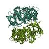 8ohwC C: citing same article ( |
|---|---|
| Similar structure data |
-Links
 PDBj
PDBj

- Assembly
| Deposited unit | 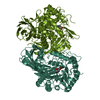
| ||||||||
|---|---|---|---|---|---|---|---|---|---|
| 1 |
| ||||||||
| Unit cell |
|
-Components
| #1: Protein | Mass: 68375.539 Da / Num. of mol.: 2 Source method: isolated from a genetically manipulated source Source: (gene. exp.) #2: Chemical |   Mass: 177.155 Da / Num. of mol.: 2 / Source method: obtained synthetically / Formula: C6H11NO5 / Feature type: SUBJECT OF INVESTIGATION Mass: 177.155 Da / Num. of mol.: 2 / Source method: obtained synthetically / Formula: C6H11NO5 / Feature type: SUBJECT OF INVESTIGATION#3: Water | ChemComp-HOH / |  Mass: 18.015 Da / Num. of mol.: 312 / Source method: isolated from a natural source / Formula: H2O Mass: 18.015 Da / Num. of mol.: 312 / Source method: isolated from a natural source / Formula: H2OHas ligand of interest | Y | |
|---|
-Experimental details
-Experiment
| Experiment | Method: X-RAY DIFFRACTION / Number of used crystals: 1 |
|---|
- Sample preparation
| Crystal | Density Matthews: 2.44 Å3/Da / Density % sol: 49.65 % |
|---|---|
| Crystal grow | Temperature: 293 K / Method: vapor diffusion, sitting drop Details: 0.1 M Bis-Tris propane pH 7.5, 20% (w/v) PEG 3350, 0.2 M NaNO3 |
-Data collection
| Diffraction | Mean temperature: 100 K / Serial crystal experiment: N |
|---|---|
| Diffraction source | Source: SYNCHROTRON / Site: Diamond / Beamline: I02 / Wavelength: 0.9795 Å |
| Detector | Type: DECTRIS PILATUS 6M / Detector: PIXEL / Date: Apr 18, 2016 |
| Radiation | Protocol: SINGLE WAVELENGTH / Monochromatic (M) / Laue (L): M / Scattering type: x-ray |
| Radiation wavelength | Wavelength: 0.9795 Å / Relative weight: 1 |
| Reflection | Resolution: 1.95→67.04 Å / Num. obs: 94487 / % possible obs: 98.4 % / Redundancy: 4.1 % / CC1/2: 0.997 / Net I/σ(I): 8.1 |
| Reflection shell | Resolution: 1.95→1.98 Å / Num. unique obs: 4628 / CC1/2: 0.717 |
- Processing
| Software |
| |||||||||||||||||||||||||||||||||||||||||||||||||||||||||||||||||||||||||||||||||||||||||||||||||||||||||||||||||||||||||||||||||||||||||||||||||||||||||||
|---|---|---|---|---|---|---|---|---|---|---|---|---|---|---|---|---|---|---|---|---|---|---|---|---|---|---|---|---|---|---|---|---|---|---|---|---|---|---|---|---|---|---|---|---|---|---|---|---|---|---|---|---|---|---|---|---|---|---|---|---|---|---|---|---|---|---|---|---|---|---|---|---|---|---|---|---|---|---|---|---|---|---|---|---|---|---|---|---|---|---|---|---|---|---|---|---|---|---|---|---|---|---|---|---|---|---|---|---|---|---|---|---|---|---|---|---|---|---|---|---|---|---|---|---|---|---|---|---|---|---|---|---|---|---|---|---|---|---|---|---|---|---|---|---|---|---|---|---|---|---|---|---|---|---|---|---|
| Refinement | Method to determine structure: MOLECULAR REPLACEMENT / Resolution: 1.95→61.835 Å / Cor.coef. Fo:Fc: 0.958 / Cor.coef. Fo:Fc free: 0.913 / WRfactor Rfree: 0.343 / WRfactor Rwork: 0.245 / SU B: 7.175 / SU ML: 0.192 / Average fsc free: 0.7935 / Average fsc work: 0.817 / Cross valid method: FREE R-VALUE / ESU R: 0.195 / ESU R Free: 0.188 Details: Hydrogens have been added in their riding positions
| |||||||||||||||||||||||||||||||||||||||||||||||||||||||||||||||||||||||||||||||||||||||||||||||||||||||||||||||||||||||||||||||||||||||||||||||||||||||||||
| Solvent computation | Ion probe radii: 0.8 Å / Shrinkage radii: 0.8 Å / VDW probe radii: 1.2 Å / Solvent model: MASK BULK SOLVENT | |||||||||||||||||||||||||||||||||||||||||||||||||||||||||||||||||||||||||||||||||||||||||||||||||||||||||||||||||||||||||||||||||||||||||||||||||||||||||||
| Displacement parameters | Biso mean: 49.837 Å2
| |||||||||||||||||||||||||||||||||||||||||||||||||||||||||||||||||||||||||||||||||||||||||||||||||||||||||||||||||||||||||||||||||||||||||||||||||||||||||||
| Refinement step | Cycle: LAST / Resolution: 1.95→61.835 Å
| |||||||||||||||||||||||||||||||||||||||||||||||||||||||||||||||||||||||||||||||||||||||||||||||||||||||||||||||||||||||||||||||||||||||||||||||||||||||||||
| Refine LS restraints |
| |||||||||||||||||||||||||||||||||||||||||||||||||||||||||||||||||||||||||||||||||||||||||||||||||||||||||||||||||||||||||||||||||||||||||||||||||||||||||||
| LS refinement shell |
|
