 Movie
Movie Controller

[English] 日本語
 Yorodumi
- PDB-8kgm: Structure of African swine fever virus topoisomerase II in comple... -
+ Open data

- Basic information
| Entry | Database: PDB / ID: 8kgm | |||||||||||||||||||||||||||||||||||||||
|---|---|---|---|---|---|---|---|---|---|---|---|---|---|---|---|---|---|---|---|---|---|---|---|---|---|---|---|---|---|---|---|---|---|---|---|---|---|---|---|---|
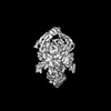
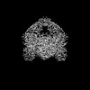
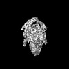
| Title | Structure of African swine fever virus topoisomerase II in complex with dsDNA | |||||||||||||||||||||||||||||||||||||||
 Components Components |
| |||||||||||||||||||||||||||||||||||||||
 Keywords Keywords | VIRAL PROTEIN/DNA / topo 2 / VIRAL PROTEIN-DNA COMPLEX | |||||||||||||||||||||||||||||||||||||||
| Function / homology |  Function and homology information Function and homology informationsister chromatid segregation / DNA topoisomerase type II (double strand cut, ATP-hydrolyzing) activity / DNA topoisomerase (ATP-hydrolysing) / DNA topological change / host cell cytoplasm / DNA binding / ATP binding / metal ion binding Similarity search - Function | |||||||||||||||||||||||||||||||||||||||
| Biological species |   African swine fever virus African swine fever virusDNA molecule (others) | |||||||||||||||||||||||||||||||||||||||
| Method | ELECTRON MICROSCOPY / single particle reconstruction / cryo EM / Resolution: 4.8 Å | |||||||||||||||||||||||||||||||||||||||
 Authors Authors | Cong, J. / Xin, Y. / Li, X. / Chen, Y. | |||||||||||||||||||||||||||||||||||||||
| Funding support | 1items
| |||||||||||||||||||||||||||||||||||||||
 Citation Citation | Journal: Nat Commun / Year: 2024 Title: Structural insights into the DNA topoisomerase II of the African swine fever virus. Authors: Jingyuan Cong / Yuhui Xin / Huiling Kang / Yunge Yang / Chenlong Wang / Dongming Zhao / Xuemei Li / Zihe Rao / Yutao Chen /  Abstract: Type II topoisomerases are ubiquitous enzymes that play a pivotal role in modulating the topological configuration of double-stranded DNA. These topoisomerases are required for DNA metabolism and ...Type II topoisomerases are ubiquitous enzymes that play a pivotal role in modulating the topological configuration of double-stranded DNA. These topoisomerases are required for DNA metabolism and have been extensively studied in both prokaryotic and eukaryotic organisms. However, our understanding of virus-encoded type II topoisomerases remains limited. One intriguing example is the African swine fever virus, which stands as the sole mammalian-infecting virus encoding a type II topoisomerase. In this work, we use several approaches including cryo-EM, X-ray crystallography, and biochemical assays to investigate the structure and function of the African swine fever virus type II topoisomerase, pP1192R. We determine the structures of pP1192R in different conformational states and confirm its enzymatic activity in vitro. Collectively, our results illustrate the basic mechanisms of viral type II topoisomerases, increasing our understanding of these enzymes and presenting a potential avenue for intervention strategies to mitigate the impact of the African swine fever virus. | |||||||||||||||||||||||||||||||||||||||
| History |
|
- Structure visualization
| Structure viewer | Molecule: MolmilJmol/JSmol |
|---|
- Downloads & links
-Download
| PDBx/mmCIF format | 8kgm.cif.gz | 629.8 KB | Display | PDBx/mmCIF format |
|---|---|---|---|---|
| PDB format | pdb8kgm.ent.gz | 509.2 KB | Display | PDB format |
| PDBx/mmJSON format | 8kgm.json.gz | Tree view | PDBx/mmJSON format | |
| Others |  Other downloads Other downloads |
-Validation report
| Arichive directory | https://data.pdbj.org/pub/pdb/validation_reports/kg/8kgmftp://data.pdbj.org/pub/pdb/validation_reports/kg/8kgm | HTTPS FTP |
|---|
-Related structure data
| Related structure data | 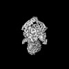 37226MC  8kglC  8kgnC  8kgoC  8kgpC  8kgqC  8kgrC 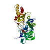 8kgsC 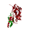 8kgtC M: map data used to model this data C: citing same article ( |
|---|---|
| Similar structure data |


-Links
 PDBj
PDBj


- Assembly
| Deposited unit | 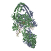
|
|---|---|
| 1 |
|
-Components
| #1: Protein | Mass: 138093.359 Da / Num. of mol.: 2 Source method: isolated from a genetically manipulated source Source: (gene. exp.) African swine fever virus / Gene: P1192R CDS / Production host:   Spodoptera frugiperda (fall armyworm) / References: UniProt: A0A2X0THW2 Spodoptera frugiperda (fall armyworm) / References: UniProt: A0A2X0THW2#2: DNA chain | | Mass: 16058.391 Da / Num. of mol.: 1 / Source method: obtained synthetically / Source: (synth.) DNA molecule (others) #3: DNA chain | | Mass: 15965.361 Da / Num. of mol.: 1 / Source method: obtained synthetically / Source: (synth.) DNA molecule (others) Has protein modification | N | |
|---|
-Experimental details
-Experiment
| Experiment | Method: ELECTRON MICROSCOPY |
|---|---|
| EM experiment | Aggregation state: PARTICLE / 3D reconstruction method: single particle reconstruction |
- Sample preparation
| Component | Name: pP1192R / Type: COMPLEX / Details: dimer / Entity ID: #1 / Source: RECOMBINANT |
|---|---|
| Source (natural) | Organism: African swine fever virus |
| Source (recombinant) | Organism: Spodoptera frugiperda (fall armyworm) |
| Buffer solution | pH: 7.2 |
| Specimen | Embedding applied: NO / Shadowing applied: NO / Staining applied: NO / Vitrification applied: YES |
| Vitrification | Cryogen name: ETHANE |
- Electron microscopy imaging
| Experimental equipment |  Model: Titan Krios / Image courtesy: FEI Company |
|---|---|
| Microscopy | Model: FEI TITAN KRIOS |
| Electron gun | Electron source:  FIELD EMISSION GUN / Accelerating voltage: 300 kV / Illumination mode: FLOOD BEAM FIELD EMISSION GUN / Accelerating voltage: 300 kV / Illumination mode: FLOOD BEAM |
| Electron lens | Mode: BRIGHT FIELD / Nominal defocus max: 2500 nm / Nominal defocus min: 1200 nm |
| Image recording | Electron dose: 60 e/Å2 / Film or detector model: GATAN K2 SUMMIT (4k x 4k) |
- Processing
| EM software | Name: PHENIX / Category: model refinement | ||||||||||||||||||||||||
|---|---|---|---|---|---|---|---|---|---|---|---|---|---|---|---|---|---|---|---|---|---|---|---|---|---|
| CTF correction | Type: PHASE FLIPPING AND AMPLITUDE CORRECTION | ||||||||||||||||||||||||
| 3D reconstruction | Resolution: 4.8 Å / Resolution method: FSC 0.143 CUT-OFF / Num. of particles: 19000 / Symmetry type: POINT | ||||||||||||||||||||||||
| Refine LS restraints |
|
