[English] 日本語
Yorodumi
- PDB-8ka5: Arabidopsis AP endonuclease ARP complex with 20bp THF-containing DNA -

+
Open data


ID or keywords:

Loading...

-
Basic information

Entry
Database: PDB / ID: 8ka5
TitleArabidopsis AP endonuclease ARP complex with 20bp THF-containing DNA
Components
  • DNA (43-MER)
  • DNA-(apurinic or apyrimidinic site) endonuclease, chloroplastic
KeywordsHYDROLASE/DNA / AP / abasic site / endonuclease / Arabidopsis / HYDROLASE-DNA complex
Function / homology
Function and homology information


chloroplast nucleoid / phosphodiesterase I activity / double-stranded DNA 3'-5' DNA exonuclease activity / exodeoxyribonuclease III / phosphatase activity / DNA-(apurinic or apyrimidinic site) endonuclease activity / 3'-5' exonuclease activity / base-excision repair / endonuclease activity / positive regulation of DNA-templated transcription ...chloroplast nucleoid / phosphodiesterase I activity / double-stranded DNA 3'-5' DNA exonuclease activity / exodeoxyribonuclease III / phosphatase activity / DNA-(apurinic or apyrimidinic site) endonuclease activity / 3'-5' exonuclease activity / base-excision repair / endonuclease activity / positive regulation of DNA-templated transcription / DNA binding / nucleus / metal ion binding
Similarity search - Function
AP endonucleases family 1 signature 2. / AP endonuclease 1, conserved site / AP endonucleases family 1 signature 3. / AP endonuclease 1, binding site / AP endonucleases family 1 signature 1. / SAP domain superfamily / AP endonuclease 1 / AP endonucleases family 1 profile. / SAP domain / SAP motif profile. ...AP endonucleases family 1 signature 2. / AP endonuclease 1, conserved site / AP endonucleases family 1 signature 3. / AP endonuclease 1, binding site / AP endonucleases family 1 signature 1. / SAP domain superfamily / AP endonuclease 1 / AP endonucleases family 1 profile. / SAP domain / SAP motif profile. / Putative DNA-binding (bihelical) motif predicted to be involved in chromosomal organisation / SAP domain / Endonuclease/exonuclease/phosphatase / Endonuclease/Exonuclease/phosphatase family / Endonuclease/exonuclease/phosphatase superfamily
Similarity search - Domain/homology
1',2'-DIDEOXYRIBOFURANOSE-5'-PHOSPHATE / DNA / DNA (> 10) / DNA-(apurinic or apyrimidinic site) endonuclease, chloroplastic
Similarity search - Component
Biological speciesArabidopsis thaliana (thale cress)
synthetic construct (others)
MethodX-RAY DIFFRACTION / SYNCHROTRON / MOLECULAR REPLACEMENT / Resolution: 2.8 Å
AuthorsGuo, W.T. / Wu, B.X.
Funding support1items
OrganizationGrant numberCountry
Not funded
CitationJournal: Structure / Year: 2024
Title: Structural insights into the catalytic mechanism of the AP endonuclease AtARP.
Authors: Guo, W. / Wu, W. / Wen, Y. / Gao, Y. / Zhuang, S. / Meng, C. / Chen, H. / Zhao, Z. / Hu, K. / Wu, B.
History
DepositionAug 2, 2023Deposition site: PDBJ / Processing site: PDBC
Revision 1.0Feb 21, 2024Provider: repository / Type: Initial release
Revision 1.1Mar 27, 2024Group: Database references / Structure summary / Category: citation / citation_author / struct
Item: _citation.country / _citation.journal_abbrev ..._citation.country / _citation.journal_abbrev / _citation.journal_id_ASTM / _citation.journal_id_CSD / _citation.journal_id_ISSN / _citation.pdbx_database_id_DOI / _citation.title / _citation.year / _struct.title
Revision 1.2Apr 3, 2024Group: Database references / Category: citation / Item: _citation.pdbx_database_id_PubMed / _citation.title
Revision 1.3Jun 19, 2024Group: Database references / Category: citation
Item: _citation.journal_volume / _citation.page_first / _citation.page_last

-
Structure visualization

Structure viewerMolecule:
MolmilJmol/JSmol

Downloads & links

-
Assembly

Deposited unit
A: DNA-(apurinic or apyrimidinic site) endonuclease, chloroplastic
B: DNA (43-MER)
C: DNA (43-MER)
D: DNA (43-MER)
hetero molecules


Theoretical massNumber of molelcules
Total (without water)73,6965
Polymers73,4984
Non-polymers1981
Water00
1


  • Idetical with deposited unit
  • defined by author&software
  • Evidence: gel filtration
TypeNameSymmetry operationNumber
identity operation1_555x,y,z1
Buried area4510 Å2
ΔGint-23 kcal/mol
Surface area17780 Å2
MethodPISA
Unit cell
Length a, b, c (Å)72.093, 72.093, 188.189
Angle α, β, γ (deg.)90.000, 90.000, 120.000
Int Tables number154
Space group name H-MP3221
Space group name HallP322"
Symmetry operation#1: x,y,z
#2: -y,x-y,z+2/3
#3: -x+y,-x,z+1/3
#4: x-y,-y,-z+1/3
#5: -x,-x+y,-z+2/3
#6: y,x,-z

-
Components

#1: Protein DNA-(apurinic or apyrimidinic site) endonuclease, chloroplastic / Apurinic endonuclease-redox protein


Mass: 33704.281 Da / Num. of mol.: 1
Source method: isolated from a genetically manipulated source
Source: (gene. exp.) Arabidopsis thaliana (thale cress) / Gene: ARP, REF, At2g41460, T26J13.5 / Production host: Escherichia coli BL21(DE3) (bacteria) / References: UniProt: P45951, exodeoxyribonuclease III
#2: DNA chain DNA (43-MER)


Mass: 13264.459 Da / Num. of mol.: 3 / Source method: obtained synthetically / Source: (synth.) synthetic construct (others)
#3: Chemical ChemComp-3DR / 1',2'-DIDEOXYRIBOFURANOSE-5'-PHOSPHATE / ABASIC DIDEOXYRIBOSE


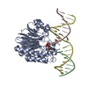
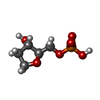
Type: DNA linking / Mass: 198.111 Da / Num. of mol.: 1 / Source method: obtained synthetically / Formula: C5H11O6P / Feature type: SUBJECT OF INVESTIGATION
Has ligand of interestY

-
Experimental details

-
Experiment

ExperimentMethod: X-RAY DIFFRACTION / Number of used crystals: 1

-
Sample preparation

CrystalDensity Matthews: 1.92 Å3/Da / Density % sol: 35.96 %
Crystal growTemperature: 293 K / Method: vapor diffusion, hanging drop
Details: 0.2 M Ammonium acetate, 0.1 M Sodium citrate tribasic dihydrate pH 5.6, 30% w/v Polyethylene glycol 4,000

-
Data collection

DiffractionMean temperature: 100 K / Serial crystal experiment: N
Diffraction sourceSource: SYNCHROTRON / Site: SSRF / Beamline: BL18U1 / Wavelength: 0.97915 Å
DetectorType: DECTRIS PILATUS3 6M / Detector: PIXEL / Date: Sep 26, 2022
RadiationProtocol: SINGLE WAVELENGTH / Monochromatic (M) / Laue (L): M / Scattering type: x-ray
Radiation wavelengthWavelength: 0.97915 Å / Relative weight: 1
ReflectionResolution: 2.8→30 Å / Num. obs: 14020 / % possible obs: 95.9 % / Redundancy: 19.3 % / Biso Wilson estimate: 79.49 Å2 / CC1/2: 0.999 / Rmerge(I) obs: 0.12 / Rpim(I) all: 0.028 / Rrim(I) all: 0.124 / Net I/σ(I): 18.8
Reflection shellResolution: 2.8→2.95 Å / Redundancy: 19.4 % / Rmerge(I) obs: 1.933 / Mean I/σ(I) obs: 2 / Num. unique obs: 2079 / CC1/2: 0.833 / Rpim(I) all: 0.444 / Rrim(I) all: 1.984 / % possible all: 99.7

-
Processing

Software
NameVersionClassification
PHENIX1.20.1_4487refinement
XDSdata reduction
SCALAdata scaling
PHASERphasing
RefinementMethod to determine structure: MOLECULAR REPLACEMENT / Resolution: 2.8→29.63 Å / SU ML: 0.4057 / Cross valid method: FREE R-VALUE / σ(F): 1.35 / Phase error: 32.141
Stereochemistry target values: GeoStd + Monomer Library + CDL v1.2
RfactorNum. reflection% reflection
Rfree0.2586 723 5.19 %
Rwork0.2014 13204 -
obs0.2043 13927 95.29 %
Solvent computationShrinkage radii: 0.9 Å / VDW probe radii: 1.1 Å / Solvent model: FLAT BULK SOLVENT MODEL
Displacement parametersBiso mean: 82.54 Å2
Refinement stepCycle: LAST / Resolution: 2.8→29.63 Å
ProteinNucleic acidLigandSolventTotal
Num. atoms2706 407 0 0 3113
Refine LS restraints
Refine-IDTypeDev idealNumber
X-RAY DIFFRACTIONf_bond_d0.00953269
X-RAY DIFFRACTIONf_angle_d1.21574611
X-RAY DIFFRACTIONf_chiral_restr0.0691509
X-RAY DIFFRACTIONf_plane_restr0.0127456
X-RAY DIFFRACTIONf_dihedral_angle_d28.0934713
LS refinement shell
Resolution (Å)Rfactor RfreeNum. reflection RfreeRfactor RworkNum. reflection RworkRefine-ID% reflection obs (%)
2.8-3.020.47121150.33452730X-RAY DIFFRACTION99.13
3.02-3.320.31091710.27212614X-RAY DIFFRACTION97.55
3.32-3.80.31341490.22812291X-RAY DIFFRACTION84.34
3.8-4.780.22641250.18812672X-RAY DIFFRACTION95.56
4.78-29.630.21871630.16622897X-RAY DIFFRACTION99.64
Refinement TLS params.Method: refined / Origin x: 19.0121392069 Å / Origin y: -17.9697739802 Å / Origin z: -10.493147253 Å
111213212223313233
T0.719331567333 Å2-0.0517287976263 Å20.0674782080066 Å2-0.416489042882 Å2-0.0514610112119 Å2--0.56779286824 Å2
L2.41254600373 °20.0023505329969 °20.903061034582 °2-1.17412977766 °2-0.342239100415 °2--2.89754940911 °2
S0.163206600209 Å °0.0395871938973 Å °0.245267514459 Å °0.238106365015 Å °-0.0214466503259 Å °0.0739170633843 Å °-0.0291732634825 Å °0.201338256089 Å °-0.106705011867 Å °
Refinement TLS groupSelection details: all

+
About Yorodumi

-
News

-
Feb 9, 2022. New format data for meta-information of EMDB entries

New format data for meta-information of EMDB entries

  • Version 3 of the EMDB header file is now the official format.
  • The previous official version 1.9 will be removed from the archive.

Related info.:EMDB header

External links:wwPDB to switch to version 3 of the EMDB data model

-
Aug 12, 2020. Covid-19 info

Covid-19 info

URL: https://pdbjlvh1.pdbj.org/emnavi/covid19.php

New page: Covid-19 featured information page in EM Navigator.

Related info.:Covid-19 info / Mar 5, 2020. Novel coronavirus structure data

+
Mar 5, 2020. Novel coronavirus structure data

Novel coronavirus structure data

Related info.:Yorodumi Speices / Aug 12, 2020. Covid-19 info

External links:COVID-19 featured content - PDBj / Molecule of the Month (242):Coronavirus Proteases

+
Jan 31, 2019. EMDB accession codes are about to change! (news from PDBe EMDB page)

EMDB accession codes are about to change! (news from PDBe EMDB page)

  • The allocation of 4 digits for EMDB accession codes will soon come to an end. Whilst these codes will remain in use, new EMDB accession codes will include an additional digit and will expand incrementally as the available range of codes is exhausted. The current 4-digit format prefixed with “EMD-” (i.e. EMD-XXXX) will advance to a 5-digit format (i.e. EMD-XXXXX), and so on. It is currently estimated that the 4-digit codes will be depleted around Spring 2019, at which point the 5-digit format will come into force.
  • The EM Navigator/Yorodumi systems omit the EMD- prefix.

Related info.:Q: What is EMD? / ID/Accession-code notation in Yorodumi/EM Navigator

External links:EMDB Accession Codes are Changing Soon! / Contact to PDBj

+
Jul 12, 2017. Major update of PDB

Major update of PDB

  • wwPDB released updated PDB data conforming to the new PDBx/mmCIF dictionary.
  • This is a major update changing the version number from 4 to 5, and with Remediation, in which all the entries are updated.
  • In this update, many items about electron microscopy experimental information are reorganized (e.g. em_software).
  • Now, EM Navigator and Yorodumi are based on the updated data.

External links:wwPDB Remediation / Enriched Model Files Conforming to OneDep Data Standards Now Available in the PDB FTP Archive

-
Yorodumi

Thousand views of thousand structures

  • Yorodumi is a browser for structure data from EMDB, PDB, SASBDB, etc.
  • This page is also the successor to EM Navigator detail page, and also detail information page/front-end page for Omokage search.
  • The word "yorodu" (or yorozu) is an old Japanese word meaning "ten thousand". "mi" (miru) is to see.

Related info.:EMDB / PDB / SASBDB / Comparison of 3 databanks / Yorodumi Search / Aug 31, 2016. New EM Navigator & Yorodumi / Yorodumi Papers / Jmol/JSmol / Function and homology information / Changes in new EM Navigator and Yorodumi

Read more