 Movie
Movie Controller


+ Open data

- Basic information
| Entry | Database: PDB / ID: 8jxs | |||||||||
|---|---|---|---|---|---|---|---|---|---|---|
| Title | Structure of nanobody-bound DRD1_PF-6142 complex | |||||||||
 Components Components |
| |||||||||
 Keywords Keywords | MEMBRANE PROTEIN / GPCR / DRD1 / PF-6142 | |||||||||
| Function / homology |  Function and homology information Function and homology informationdopamine neurotransmitter receptor activity, coupled via Gs / dopamine neurotransmitter receptor activity / cerebral cortex GABAergic interneuron migration / Dopamine receptors / regulation of dopamine uptake involved in synaptic transmission / dopamine binding / phospholipase C-activating dopamine receptor signaling pathway / heterotrimeric G-protein binding / 9+0 non-motile cilium / G protein-coupled receptor complex ...dopamine neurotransmitter receptor activity, coupled via Gs / dopamine neurotransmitter receptor activity / cerebral cortex GABAergic interneuron migration / Dopamine receptors / regulation of dopamine uptake involved in synaptic transmission / dopamine binding / phospholipase C-activating dopamine receptor signaling pathway / heterotrimeric G-protein binding / 9+0 non-motile cilium / G protein-coupled receptor complex / modification of postsynaptic structure / adult walking behavior / positive regulation of potassium ion transport / arrestin family protein binding / non-motile cilium / G protein-coupled dopamine receptor signaling pathway / IgG binding / mating behavior / ciliary membrane / dopamine metabolic process / detection of maltose stimulus / transmission of nerve impulse / carbohydrate transport / carbohydrate transmembrane transporter activity / maltose binding / G-protein alpha-subunit binding / maltose transport / maltodextrin transmembrane transport / G protein-coupled receptor signaling pathway, coupled to cyclic nucleotide second messenger / ATP-binding cassette (ABC) transporter complex, substrate-binding subunit-containing / adenylate cyclase-activating adrenergic receptor signaling pathway / positive regulation of synaptic transmission, glutamatergic / synapse assembly / presynaptic modulation of chemical synaptic transmission / visual learning / ATP-binding cassette (ABC) transporter complex / positive regulation of release of sequestered calcium ion into cytosol / cell chemotaxis / vasodilation / GABA-ergic synapse / G protein-coupled receptor activity / cilium / cellular response to catecholamine stimulus / adenylate cyclase-activating dopamine receptor signaling pathway / outer membrane-bounded periplasmic space / adenylate cyclase-activating G protein-coupled receptor signaling pathway / presynaptic membrane / dendritic spine / G alpha (s) signalling events / positive regulation of MAPK cascade / periplasmic space / postsynaptic membrane / response to xenobiotic stimulus / positive regulation of cell migration / DNA damage response / endoplasmic reticulum membrane / glutamatergic synapse / membrane / nucleus / plasma membrane Similarity search - Function | |||||||||
| Biological species |  Homo sapiens (human) Homo sapiens (human)  Escherichia coli K-12 (bacteria) Escherichia coli K-12 (bacteria) Staphylococcus aureus (bacteria) Staphylococcus aureus (bacteria)  Mus musculus (house mouse) Mus musculus (house mouse) | |||||||||
| Method | ELECTRON MICROSCOPY / single particle reconstruction / cryo EM / Resolution: 3 Å | |||||||||
 Authors Authors | Zhuang, Y. / Xu, Y. / Fan, L. / Wang, S. / Xu, H.E. | |||||||||
| Funding support |  China, 2items China, 2items
| |||||||||
 Citation Citation | Journal: Neuron / Year: 2024 Title: Structural basis of psychedelic LSD recognition at dopamine D receptor. Authors: Luyu Fan / Youwen Zhuang / Hongyu Wu / Huiqiong Li / Youwei Xu / Yue Wang / Licong He / Shishan Wang / Zhangcheng Chen / Jianjun Cheng / H Eric Xu / Sheng Wang / Abstract: Understanding the kinetics of LSD in receptors and subsequent induced signaling is crucial for comprehending both the psychoactive and therapeutic effects of LSD. Despite extensive research on LSD's ...Understanding the kinetics of LSD in receptors and subsequent induced signaling is crucial for comprehending both the psychoactive and therapeutic effects of LSD. Despite extensive research on LSD's interactions with serotonin 2A and 2B receptors, its behavior on other targets, including dopamine receptors, has remained elusive. Here, we present cryo-EM structures of LSD/PF6142-bound dopamine D receptor (DRD1)-legobody complexes, accompanied by a β-arrestin-mimicking nanobody, NBA3, shedding light on the determinants of G protein coupling versus β-arrestin coupling. Structural analysis unveils a distinctive binding mode of LSD in DRD1, particularly with the ergoline moiety oriented toward TM4. Kinetic investigations uncover an exceptionally rapid dissociation rate of LSD in DRD1, attributed to the flexibility of extracellular loop 2 (ECL2). Moreover, G protein can stabilize ECL2 conformation, leading to a significant slowdown in ligand's dissociation rate. These findings establish a solid foundation for further exploration of G protein-coupled receptor (GPCR) dynamics and their relevance to signal transduction. | |||||||||
| History |
|
- Structure visualization
| Structure viewer | Molecule: MolmilJmol/JSmol |
|---|
- Downloads & links
-Download
| PDBx/mmCIF format | 8jxs.cif.gz | 226.4 KB | Display | PDBx/mmCIF format |
|---|---|---|---|---|
| PDB format | pdb8jxs.ent.gz | 170.4 KB | Display | PDB format |
| PDBx/mmJSON format | 8jxs.json.gz | Tree view | PDBx/mmJSON format | |
| Others |  Other downloads Other downloads |
-Validation report
| Arichive directory | https://data.pdbj.org/pub/pdb/validation_reports/jx/8jxsftp://data.pdbj.org/pub/pdb/validation_reports/jx/8jxs | HTTPS FTP |
|---|
-Related structure data
| Related structure data |  36711MC  8jxrC M: map data used to model this data C: citing same article ( |
|---|---|
| Similar structure data |

-Links
 PDBj
PDBj








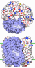


- Assembly
| Deposited unit | 
|
|---|---|
| 1 |
|
-Components
-Antibody , 4 types, 4 molecules BCHL
| #2: Antibody | Mass: 13493.983 Da / Num. of mol.: 1 Source method: isolated from a genetically manipulated source Source: (gene. exp.) Homo sapiens (human) / Production host:   Spodoptera frugiperda (fall armyworm) Spodoptera frugiperda (fall armyworm) |
|---|---|
| #3: Antibody | Mass: 60575.715 Da / Num. of mol.: 1 / Mutation: E360Q,K363A,D364F,T367I,R368L,D404E,A405H Source method: isolated from a genetically manipulated source Source: (gene. exp.) Escherichia coli K-12 (bacteria), (gene. exp.) Staphylococcus aureus (bacteria)Gene: malE, spa, spa, SAV0111, spg / Production host: Spodoptera frugiperda (fall armyworm)References: UniProt: P0AEX9, UniProt: P38507, UniProt: P0A015, UniProt: P06654 |
| #4: Antibody | Mass: 27396.742 Da / Num. of mol.: 1 Source method: isolated from a genetically manipulated source Source: (gene. exp.) Mus musculus (house mouse) / Production host: Spodoptera frugiperda (fall armyworm) |
| #5: Antibody | Mass: 26317.523 Da / Num. of mol.: 1 Source method: isolated from a genetically manipulated source Source: (gene. exp.) Mus musculus (house mouse) / Production host: Spodoptera frugiperda (fall armyworm) |
-Protein / Non-polymers , 2 types, 2 molecules A
| #1: Protein | Mass: 39187.793 Da / Num. of mol.: 1 / Mutation: L112W,S325A Source method: isolated from a genetically manipulated source Source: (gene. exp.) Homo sapiens (human) / Gene: DRD1 / Production host: Spodoptera frugiperda (fall armyworm) / References: UniProt: P21728 |
|---|---|
| #6: Chemical | ChemComp-V6X / Mass: 356.377 Da / Num. of mol.: 1 / Source method: obtained synthetically / Formula: C21H16N4O2 |
-Details
| Has ligand of interest | Y |
|---|---|
| Has protein modification | Y |
-Experimental details
-Experiment
| Experiment | Method: ELECTRON MICROSCOPY |
|---|---|
| EM experiment | Aggregation state: PARTICLE / 3D reconstruction method: single particle reconstruction |
- Sample preparation
| Component |
| ||||||||||||||||||||||||||||||
|---|---|---|---|---|---|---|---|---|---|---|---|---|---|---|---|---|---|---|---|---|---|---|---|---|---|---|---|---|---|---|---|
| Molecular weight | Value: 0.17 MDa / Experimental value: NO | ||||||||||||||||||||||||||||||
| Source (natural) |
| ||||||||||||||||||||||||||||||
| Source (recombinant) | Organism: Spodoptera frugiperda (fall armyworm) | ||||||||||||||||||||||||||||||
| Buffer solution | pH: 7.5 | ||||||||||||||||||||||||||||||
| Specimen | Embedding applied: NO / Shadowing applied: NO / Staining applied: NO / Vitrification applied: YES | ||||||||||||||||||||||||||||||
| Vitrification | Cryogen name: ETHANE |
- Electron microscopy imaging
| Experimental equipment |  Model: Titan Krios / Image courtesy: FEI Company |
|---|---|
| Microscopy | Model: FEI TITAN KRIOS |
| Electron gun | Electron source:  FIELD EMISSION GUN / Accelerating voltage: 300 kV / Illumination mode: FLOOD BEAM FIELD EMISSION GUN / Accelerating voltage: 300 kV / Illumination mode: FLOOD BEAM |
| Electron lens | Mode: BRIGHT FIELD / Nominal defocus max: 5000 nm / Nominal defocus min: 1200 nm |
| Image recording | Electron dose: 50 e/Å2 / Film or detector model: GATAN K3 (6k x 4k) |
- Processing
| CTF correction | Type: NONE |
|---|---|
| 3D reconstruction | Resolution: 3 Å / Resolution method: FSC 0.143 CUT-OFF / Num. of particles: 104462 / Symmetry type: POINT |
