 Movie
Movie Controller

[English] 日本語
 Yorodumi
- PDB-8jpd: Focused refinement structure of GRK2 in NTSR1-GRK2-Galpha(q) complexes -
+ Open data

- Basic information
| Entry | Database: PDB / ID: 8jpd | ||||||
|---|---|---|---|---|---|---|---|
| Title | Focused refinement structure of GRK2 in NTSR1-GRK2-Galpha(q) complexes | ||||||
 Components Components | Beta-adrenergic receptor kinase 1 | ||||||
 Keywords Keywords | SIGNALING PROTEIN / Biased signaling | ||||||
| Function / homology |  Function and homology information Function and homology informationCalmodulin induced events / negative regulation of the force of heart contraction by chemical signal / negative regulation of relaxation of smooth muscle / beta-adrenergic-receptor kinase / Edg-2 lysophosphatidic acid receptor binding / Activation of SMO / alpha-2A adrenergic receptor binding / beta-adrenergic receptor kinase activity / G-protein-coupled receptor kinase / G protein-coupled receptor kinase activity ...Calmodulin induced events / negative regulation of the force of heart contraction by chemical signal / negative regulation of relaxation of smooth muscle / beta-adrenergic-receptor kinase / Edg-2 lysophosphatidic acid receptor binding / Activation of SMO / alpha-2A adrenergic receptor binding / beta-adrenergic receptor kinase activity / G-protein-coupled receptor kinase / G protein-coupled receptor kinase activity / tachykinin receptor signaling pathway / negative regulation of striated muscle contraction / Cargo recognition for clathrin-mediated endocytosis / positive regulation of protein localization to cilium / cytoplasmic side of mitochondrial outer membrane / regulation of the force of heart contraction / desensitization of G protein-coupled receptor signaling pathway / positive regulation of smoothened signaling pathway / G protein-coupled receptor internalization / G alpha (s) signalling events / G alpha (q) signalling events / regulation of signal transduction / adenylate cyclase-activating adrenergic receptor signaling pathway / cardiac muscle contraction / viral genome replication / intracellular protein transport / G protein-coupled receptor binding / G protein-coupled acetylcholine receptor signaling pathway / presynapse / protein kinase activity / postsynapse / G protein-coupled receptor signaling pathway / symbiont entry into host cell / ATP binding / membrane / plasma membrane / cytosol / cytoplasm Similarity search - Function | ||||||
| Biological species |  | ||||||
| Method | ELECTRON MICROSCOPY / single particle reconstruction / cryo EM / Resolution: 2.81 Å | ||||||
 Authors Authors | Duan, J. / Liu, H. / Zhao, F. / Yuan, Q. / Ji, Y. / Xu, H.E. | ||||||
| Funding support |  China, 1items China, 1items
| ||||||
 Citation Citation | Journal: Nature / Year: 2023 Title: GPCR activation and GRK2 assembly by a biased intracellular agonist. Authors: Jia Duan / Heng Liu / Fenghui Zhao / Qingning Yuan / Yujie Ji / Xiaoqing Cai / Xinheng He / Xinzhu Li / Junrui Li / Kai Wu / Tianyu Gao / Shengnan Zhu / Shi Lin / Ming-Wei Wang / Xi Cheng / ...Authors: Jia Duan / Heng Liu / Fenghui Zhao / Qingning Yuan / Yujie Ji / Xiaoqing Cai / Xinheng He / Xinzhu Li / Junrui Li / Kai Wu / Tianyu Gao / Shengnan Zhu / Shi Lin / Ming-Wei Wang / Xi Cheng / Wanchao Yin / Yi Jiang / Dehua Yang / H Eric Xu / Abstract: Phosphorylation of G-protein-coupled receptors (GPCRs) by GPCR kinases (GRKs) desensitizes G-protein signalling and promotes arrestin signalling, which is also modulated by biased ligands. The ...Phosphorylation of G-protein-coupled receptors (GPCRs) by GPCR kinases (GRKs) desensitizes G-protein signalling and promotes arrestin signalling, which is also modulated by biased ligands. The molecular assembly of GRKs on GPCRs and the basis of GRK-mediated biased signalling remain largely unknown owing to the weak GPCR-GRK interactions. Here we report the complex structure of neurotensin receptor 1 (NTSR1) bound to GRK2, Gα and the arrestin-biased ligand SBI-553. The density map reveals the arrangement of the intact GRK2 with the receptor, with the N-terminal helix of GRK2 docking into the open cytoplasmic pocket formed by the outward movement of the receptor transmembrane helix 6, analogous to the binding of the G protein to the receptor. SBI-553 binds at the interface between GRK2 and NTSR1 to enhance GRK2 binding. The binding mode of SBI-553 is compatible with arrestin binding but clashes with the binding of Gα protein, thus providing a mechanism for its arrestin-biased signalling capability. In sum, our structure provides a rational model for understanding the details of GPCR-GRK interactions and GRK2-mediated biased signalling. | ||||||
| History |
|
- Structure visualization
| Structure viewer | Molecule: MolmilJmol/JSmol |
|---|
- Downloads & links
-Download
| PDBx/mmCIF format | 8jpd.cif.gz | 129.2 KB | Display | PDBx/mmCIF format |
|---|---|---|---|---|
| PDB format | pdb8jpd.ent.gz | 97.7 KB | Display | PDB format |
| PDBx/mmJSON format | 8jpd.json.gz | Tree view | PDBx/mmJSON format | |
| Others |  Other downloads Other downloads |
-Validation report
| Arichive directory | https://data.pdbj.org/pub/pdb/validation_reports/jp/8jpdftp://data.pdbj.org/pub/pdb/validation_reports/jp/8jpd | HTTPS FTP |
|---|
-Related structure data
| Related structure data |  36476MC  8jpbC  8jpcC 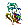 8jpeC  8jpfC M: map data used to model this data C: citing same article ( |
|---|---|
| Similar structure data |


-Links
 PDBj
PDBj


- Assembly
| Deposited unit | 
|
|---|---|
| 1 |
|
-Components
| #1: Protein | Mass: 79644.727 Da / Num. of mol.: 1 / Mutation: A292P,R295I,S455D Source method: isolated from a genetically manipulated source Source: (gene. exp.)   Spodoptera frugiperda (fall armyworm) Spodoptera frugiperda (fall armyworm)References: UniProt: P21146, beta-adrenergic-receptor kinase |
|---|---|
| #2: Chemical | ChemComp-STU /   Mass: 466.531 Da / Num. of mol.: 1 / Source method: obtained synthetically / Formula: C28H26N4O3 / Feature type: SUBJECT OF INVESTIGATION / Comment: anticancer, antifungal, antibiotic, alkaloid*YM Mass: 466.531 Da / Num. of mol.: 1 / Source method: obtained synthetically / Formula: C28H26N4O3 / Feature type: SUBJECT OF INVESTIGATION / Comment: anticancer, antifungal, antibiotic, alkaloid*YM |
| Has ligand of interest | Y |
-Experimental details
-Experiment
| Experiment | Method: ELECTRON MICROSCOPY |
|---|---|
| EM experiment | Aggregation state: PARTICLE / 3D reconstruction method: single particle reconstruction |
- Sample preparation
| Component | Name: Focused refinement structure of GRK2 in NTSR1-GRK2-Galpha(q) complexes Type: COMPLEX / Entity ID: #1 / Source: RECOMBINANT |
|---|---|
| Source (natural) | Organism: |
| Source (recombinant) | Organism: Spodoptera frugiperda (fall armyworm) |
| Buffer solution | pH: 7.4 |
| Specimen | Embedding applied: NO / Shadowing applied: NO / Staining applied: NO / Vitrification applied: YES |
| Vitrification | Cryogen name: ETHANE |
- Electron microscopy imaging
| Experimental equipment |  Model: Titan Krios / Image courtesy: FEI Company |
|---|---|
| Microscopy | Model: FEI TITAN KRIOS |
| Electron gun | Electron source:  FIELD EMISSION GUN / Accelerating voltage: 300 kV / Illumination mode: FLOOD BEAM FIELD EMISSION GUN / Accelerating voltage: 300 kV / Illumination mode: FLOOD BEAM |
| Electron lens | Mode: DARK FIELD / Nominal defocus max: 2200 nm / Nominal defocus min: 1200 nm |
| Image recording | Electron dose: 50 e/Å2 / Film or detector model: GATAN K3 BIOQUANTUM (6k x 4k) |
- Processing
| CTF correction | Type: NONE |
|---|---|
| 3D reconstruction | Resolution: 2.81 Å / Resolution method: FSC 0.143 CUT-OFF / Num. of particles: 474232 / Symmetry type: POINT |
